THE INSULIN-IGF-1 AXIS IN PMOS
The Engine Room: Hyperinsulinemia as the Central Driver of Polyendocrine Metabolic Ovarian Syndrome

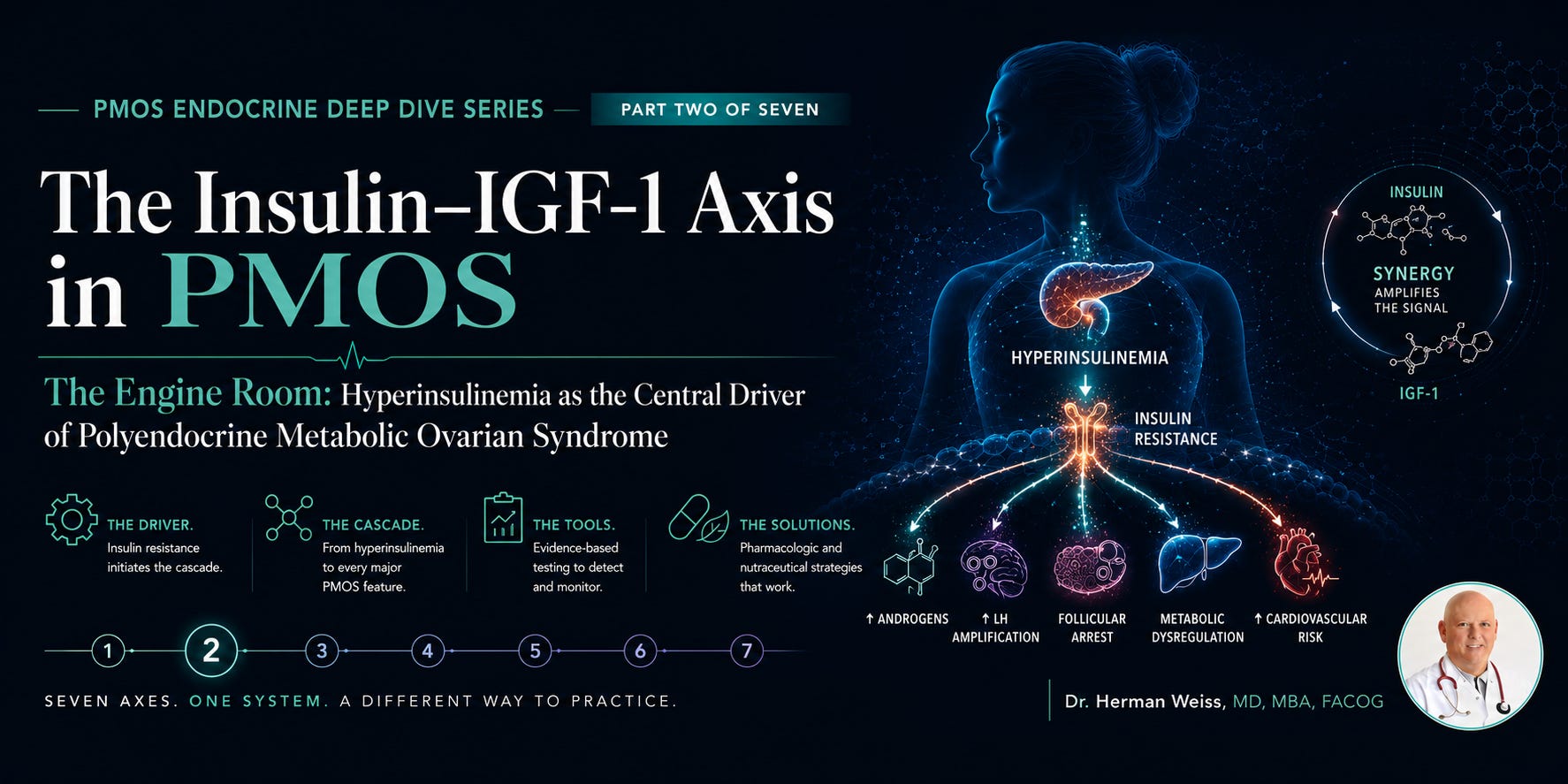
The Engine Room
In Part One, we examined the thyroid. A meaningful contributing axis in PMOS, an amplifier of the phenotype, a source of genuine diagnostic and therapeutic nuance. But the thyroid is not the engine. The thyroid is not what drives the hyperandrogenism, the anovulation, the follicular arrest, the visceral adiposity, the dyslipidemia, or the lifelong cardiovascular risk that defines this disease.
Insulin does. More precisely: chronic compensatory hyperinsulinemia, the physiological consequence of insulin resistance, is the central pathophysiological driver of PMOS. Everything else in this series is downstream of this axis. The HPO dysregulation, the androgen excess, the HPA involvement, the prolactin perturbations, all of it. When we renamed this condition Polyendocrine Metabolic Ovarian Syndrome, “metabolic” was not an adjective. It was a diagnosis.
This installment will do four things: establish the basic science of insulin signaling and why it goes wrong in PMOS; trace the pathophysiological cascade from insulin resistance to every major clinical feature; assess the laboratory tools available to diagnose and monitor the metabolic axis; and deliver an honest, referenced review of the pharmacological and nutraceutical landscape, including what genuinely works, what is overhyped, and what the current evidence actually supports.
I will not soften the findings in either direction. Some supplements here have RCT-level evidence. Some widely used interventions do not. And at least one, metformin, belongs in a separate category entirely.
1. The Biology of Insulin Resistance in PMOS: What We Know and Why It Matters
1a. Normal Insulin Signaling — The Reference Point
Insulin binds to the insulin receptor (IR), a transmembrane tyrosine kinase, triggering autophosphorylation and activation of insulin receptor substrate proteins (IRS-1, IRS-2). This activates two major downstream cascades: the PI3K-Akt-mTOR pathway (responsible for metabolic effects: glucose uptake via GLUT4 translocation, glycogen synthesis, lipogenesis) and the Ras-MAPK pathway (responsible for mitogenic and proliferative effects: cell growth, differentiation, steroidogenesis).¹
In the insulin-resistant state, the PI3K-Akt pathway becomes selectively impaired; glucose uptake is reduced and the liver, muscle, and adipose tissue become progressively resistant to insulin’s metabolic actions. The pancreatic beta cell compensates with increased insulin secretion. And here is the critical, underappreciated point:
The Central Paradox of Insulin Resistance in PMOS
The PI3K-Akt (metabolic) pathway is impaired. Glucose uptake is reduced.
The Ras-MAPK (mitogenic/steroidogenic) pathway remains fully or even hypersensitively active.
Compensatory hyperinsulinemia floods the system, driving steroidogenesis, LH amplification, and follicular arrest through the intact pathway while the metabolic derangement worsens.
The ovary, the adrenal gland, and the pituitary are not insulin-resistant in the same way peripheral tissues are. They receive the full steroidogenic and mitogenic signal from chronically elevated insulin.
This pathway selectivity is why hyperinsulinemia causes hyperandrogenism even when glucose homeostasis appears relatively preserved.
This selective insulin resistance, first characterized by Dunaif et al. in seminal work at Mount Sinai across the 1980s–1990s, remains the most important mechanistic insight in the biology of what we now call PMOS.² It explains why a woman can have a fasting glucose of 88 mg/dL and still be driving profound ovarian androgen excess through hyperinsulinemia. It explains why the ovary is a victim of a systemic metabolic disorder rather than the source of an intrinsic reproductive defect.
1b. The Molecular Defect: What Causes Insulin Resistance in PMOS?
Insulin resistance in PMOS is not simply obesity-related, although obesity amplifies it. A substantial body of evidence demonstrates an intrinsic, obesity-independent defect in insulin signaling in PMOS patients, present in lean PMOS patients and in first-degree relatives without PMOS, suggesting a primary genetic or epigenetic vulnerability.
The primary molecular lesion identified is constitutive serine phosphorylation of IRS-1 (at Ser³²³ and Ser⁷³²), mediated at least in part by an intrinsic serine kinase abnormality. Serine phosphorylation of IRS-1 inhibits its normal tyrosine phosphorylation, impairing downstream PI3K-Akt activation.³ This same serine kinase appears to phosphorylate CYP17A1, the rate-limiting enzyme in androgen biosynthesis, enhancing its activity. So the same molecular defect that drives insulin resistance simultaneously upregulates androgen synthesis. This is not a coincidence. This is shared pathophysiology.
Additional contributing mechanisms include: impaired GLUT4 expression and translocation in adipose and muscle;⁴ mitochondrial dysfunction with reduced oxidative phosphorylation capacity;⁵ elevated free fatty acids (from adipose tissue lipolysis, itself driven by insulin resistance) that activate IKKβ/NF-κB inflammatory pathways and further impair IRS-1 signaling;⁶ and dysregulated adipokine secretion reduced adiponectin, elevated leptin and resistin creating an endocrine environment that perpetuates insulin resistance from the fat depot itself.
1c. The IGF-1 Axis: The Amplifier
Insulin-like growth factor 1 (IGF-1) is structurally homologous to insulin and binds with lower affinity to the insulin receptor, as well as to its own IGF-1 receptor (IGF-1R). In the PMOS ovary, IGF-1 acts as a potent co-gonadotropin, synergizing with LH to amplify thecal androgen production and with FSH to support granulosa cell function.⁷
In hyperinsulinemia, insulin suppresses hepatic synthesis of insulin-like growth factor binding protein 1 (IGFBP-1), which normally sequesters IGF-1 in the circulation. Reduced IGFBP-1 means elevated free IGF-1 bioavailability.⁸ This amplifies ovarian androgen production beyond what insulin alone would drive, creates a permissive environment for LH-driven thecal cell proliferation, and further impairs follicular maturation by disrupting the granulosa-thecal balance needed for normal folliculogenesis.
The insulin-IGF-1 axis is therefore not a single pathway but a dual-input system, both arms of which are dysregulated in PMOS and both of which converge on the same downstream target: androgen excess and follicular arrest.

2. From Insulin Resistance to Every Clinical Feature: The Full Cascade
The power of the insulin-IGF-1 axis as an explanatory framework lies in its ability to account for every major phenotypic feature of PMOS through mechanistic pathways that are individually documented and collectively coherent. This is not a just-so story. This is traceable, step-by-step biochemistry.
Chronic hyperinsulinemia (compensatory response to peripheral insulin resistance)
→ drives downstream through two intact pathways:
Insulin directly stimulates ovarian theca cell CYP17A1 → elevated androstenedione → testosterone excess
→ simultaneously:
Insulin suppresses IGFBP-1 → elevated free IGF-1 → co-stimulates thecal androgen synthesis with LH
→ and:
LH pulse amplitude increased (via GnRH sensitization by androgens and insulin) → further thecal stimulation
→ resulting in:
Hyperandrogenism (free testosterone, androstenedione, DHEA-S elevation)
→ which causes:
Follicular arrest at preantral/antral stage, anovulation, oligomenorrhea
→ which produces the visible sign:
Polycystic-appearing ovaries on ultrasound (the morphological epiphenomenon, not the cause)
Androgen Excess: The Direct Ovarian Pathway
Nestler et al. demonstrated in a landmark 1998 New England Journal of Medicine study that reducing insulin levels with metformin significantly reduced androgen production in PCOS patients, providing direct experimental evidence for the insulin-androgen causal link.⁹ Subsequent studies with insulin-sensitizing agents across multiple drug classes have consistently reproduced this finding: lower insulin, lower androgens. This is now as close to a proven causal pathway as endocrinology offers.
SHBG: The Indirect Amplifier
Sex hormone-binding globulin (SHBG) is synthesized in the liver. Insulin suppresses hepatic SHBG synthesis through a well-characterized mechanism involving the FOXO1 transcription factor — the same factor that mediates gluconeogenesis regulation.¹⁰ Hyperinsulinemia therefore reduces SHBG, increasing the free androgen fraction even without any increase in total testosterone production. A PMOS patient with a total testosterone in the upper-normal range may have markedly elevated free testosterone simply because her SHBG is suppressed. This is why free androgen index or calculated free testosterone must be part of the androgen assessment — total testosterone alone understates the clinical picture in hyperinsulinemic patients.
Follicular Arrest and Anovulation: The Downstream Consequence
Normal follicular development requires a precise FSH-to-LH ratio and an estrogen-dominant intrafollicular milieu. In PMOS: insulin and IGF-1 impair granulosa cell FSH responsiveness through downregulation of FSH receptor expression;¹¹ the LH pulse frequency increase (driven by GnRH hypersensitization from excess androgens and direct insulin effect on the hypothalamus) shifts the LH:FSH ratio toward LH dominance; and the androgen excess itself induces granulosa cell apoptosis while stimulating thecal cell proliferation. The net result is follicles that arrest at 2–8 mm, fail to select a dominant follicle, and produce the characteristic “string of pearls” appearance on ultrasound.
These are not separate pathways. They are one amplifying loop, initiated in the insulin-IGF-1 axis, expressed in the HPO axis, and visible in the ovary. The ovary is the readout. Insulin resistance is the program.
Adipose Tissue and the Metabolic Feedforward Loop
Visceral adiposity — disproportionately elevated in PMOS even in lean patients relative to BMI-matched controls — is both a consequence and an amplifier of insulin resistance. Visceral adipocytes are metabolically active endocrine cells. They secrete: free fatty acids (impairing hepatic insulin sensitivity and promoting atherogenic dyslipidemia); resistin and TNF-α (directly impairing IRS-1 phosphorylation); and adiponectin at reduced levels (adiponectin is an insulin-sensitizer — its reduction is a marker of metabolic dysfunction and a driver of further resistance).¹²
This creates a feedforward loop: insulin resistance promotes visceral fat deposition, visceral fat worsens insulin resistance, which further promotes fat deposition. Weight gain in PMOS is not a matter of willpower or caloric excess alone — it is a metabolic architecture problem that cannot be addressed by caloric restriction in isolation.
3. Laboratory Assessment of the Insulin-IGF-1 Axis in PMOS: What to Measure and Why
This is a practical section. I want to be clear about what the evidence supports for clinical use, what is emerging, and what is not yet ready for routine clinical application.
Tier 1: What Every PMOS Patient Should Have
Fasting Insulin and Fasting Glucose
Fasting glucose alone is an inadequate screen for insulin resistance in PMOS. A patient can maintain near-normal fasting glucose through massive compensatory hyperinsulinemia for years before glucose homeostasis visibly fails. Fasting insulin must be measured alongside fasting glucose. A fasting insulin above 12–15 mIU/L in a fasting state is a meaningful signal even with normal glucose.
HOMA-IR (Homeostatic Model Assessment of Insulin Resistance)
HOMA-IR = (Fasting insulin [mIU/L] × Fasting glucose [mmol/L]) / 22.5. A value above 2.0 is generally considered elevated; above 2.5–3.0 suggests clinically significant insulin resistance in most populations. HOMA-IR is imperfect — it does not capture postprandial insulin dynamics and underestimates resistance in patients with significant hepatic insulin resistance — but it is validated, reproducible, and easily obtained from standard fasting labs.¹³ Every PMOS patient should have a HOMA-IR calculated.
Hemoglobin A1c
HbA1c captures 90-day average glucose and stratifies cardiometabolic risk. In PMOS, it serves as both a diagnostic and monitoring tool. An HbA1c of 5.7–6.4% (prediabetes range) should trigger aggressive metabolic intervention. The lifetime risk of type 2 diabetes in PCOS/PMOS is approximately 5–7 times the general population risk.
Lipid Panel
PMOS-associated dyslipidemia is specific: elevated triglycerides, reduced HDL, elevated small dense LDL particles (even with normal total LDL). A standard fasting lipid panel is essential. Where available, an LDL particle size assessment (LDL-P by NMR) adds clinically relevant information regarding atherogenic risk that total LDL misses.
Tier 2: Clinically Valuable, Evidence-Supported
2-Hour Oral Glucose Tolerance Test (OGTT) with Insulin
The OGTT with concurrent insulin levels is the most sensitive clinical tool for identifying insulin resistance and impaired glucose tolerance in PMOS. It captures the postprandial hyperinsulinemic response that HOMA-IR misses. A 2016 Endocrine Society position statement on PCOS recommended OGTT as the preferred glycemic screening method over HbA1c alone in this population, noting that HbA1c alone may miss up to 20% of impaired glucose tolerance cases.¹⁴ This is the gold standard screen for metabolic risk stratification in PMOS. Many practices do not offer it routinely. This should change.
Fasting Free Fatty Acids
Elevated fasting free fatty acids reflect adipose tissue insulin resistance and predict hepatic insulin resistance independent of BMI. Less widely available but valuable in metabolically complex patients. Increasingly included in comprehensive metabolic panels at academic centers.
Triglyceride-to-HDL Ratio
A TG:HDL ratio above 3.0 (using mg/dL units) or above 1.3 (using mmol/L) is a validated surrogate marker for insulin resistance and correlates well with hyperinsulinemia in multiple population studies.¹⁵ It is calculable from any standard lipid panel and costs nothing extra. In my practice, a TG:HDL ratio above 2.5 in a PMOS patient is treated as a metabolic red flag regardless of fasting glucose.
Tier 3: Emerging and Specialized — Not Yet Routine
IGF-1 and IGFBP-1
Measurement of serum IGF-1 and IGFBP-1 provides direct insight into the IGF axis dysregulation described above. Reduced IGFBP-1 in the presence of elevated free IGF-1 correlates with hyperinsulinemia and predicts ovarian androgen excess independent of serum insulin levels. Available at most reference laboratories; not yet part of standard PMOS guidelines but mechanistically justified.
Adiponectin
Adiponectin is an adipokine with direct insulin-sensitizing, anti-inflammatory, and anti-atherogenic properties. It is consistently reduced in PMOS patients and inversely correlates with insulin resistance severity. Low adiponectin predicts progression to type 2 diabetes independent of BMI and is now under evaluation as a therapeutic target. Not yet a routine clinical test but valuable in research-oriented or complex clinical settings.
Metabolic Lab Panel for Every PMOS Patient — Recommended BaselineFasting glucose + fasting insulin (calculate HOMA-IR)
Hemoglobin A1c
Fasting lipid panel (total cholesterol, LDL, HDL, triglycerides)
Calculate TG:HDL ratio — flag if >2.5
Liver enzymes (AST, ALT) — screen for NAFLD/MASLD, prevalent in PMOS at ~35%
Consider 2-hour OGTT with insulin levels for full glycemic risk stratification
Reassess metabolic panel every 12 months minimum — every 6 months if abnormal
4. The Insulin-Sensitizing Landscape: Pharmacology, Nutraceuticals, and the Evidence
This section covers more clinical ground than the thyroid supplement review, because the insulin axis has attracted a much larger body of evidence — including genuine RCT and meta-analysis data for several interventions. I will review each major category: established pharmacotherapy, evidence-supported nutraceuticals, overhyped supplements, and lifestyle — which belongs in this section because it is the most evidence-supported intervention of all.
The Pharmacological Foundation: Metformin
Metformin deserves its own section, not because it is a supplement, but because it sits at the center of PMOS pharmacotherapy and because the stigma around its “diabetes drug” label has directly harmed patients — as I described in the post that preceded this series. Every pharmacist who has questioned a metformin prescription for a non-diabetic PMOS patient is a product of the nomenclature failure that PMOS is designed to correct.
Metformin (biguanide class) works primarily by activating AMP-activated protein kinase (AMPK) in the liver, reducing hepatic glucose production. It also reduces intestinal glucose absorption and modestly improves peripheral insulin sensitivity. In PMOS, its downstream effects include: reduced fasting and postprandial insulin; increased SHBG (via restoration of FOXO1-mediated hepatic SHBG synthesis); reduced androgen production (direct effect on CYP17A1 via AMPK activation); and improved ovulatory function.¹⁶
The evidence base is substantial. A 2012 Cochrane review of 44 RCTs found metformin significantly improved clinical pregnancy rates and ovulation rates compared to placebo in PCOS.¹⁷ A 2023 meta-analysis in Lancet Diabetes & Endocrinology confirmed that metformin reduces androgen levels, improves menstrual regularity, and reduces metabolic risk markers in PCOS independent of weight loss.¹⁸ The evidence for metformin in PMOS is as strong as for any pharmacological intervention in reproductive endocrinology. It is the current standard of care for metabolic management in PMOS patients — full stop.
On GLP-1 receptor agonists: Semaglutide, liraglutide, and the dual GIP/GLP-1 agonist tirzepatide are generating significant interest in PMOS given their profound effects on insulin sensitivity, weight reduction, and potentially direct ovarian effects. Early data is promising: a 2023 RCT of semaglutide in PCOS showed significant reductions in BMI, testosterone, and HOMA-IR, with improved menstrual regularity.¹⁹ These agents will be addressed in full in a forthcoming supplement to this series. For now: they represent a legitimate and rapidly evolving therapeutic category that belongs in the conversation for metabolically complex PMOS patients, but their long-term reproductive safety profile is still being established.
Evidence Summary Table: Insulin-Sensitizing Interventions in PMOS
AMPK activation, reduced hepatic glucose output, reduced CYP17A1 activity
A
Clinical Reality: Gold standard pharmacotherapy. Reduces androgens, improves ovulation, reduces metabolic risk. Not a “diabetes drug” — it is a PMOS drug.
Supplement / Drug: Inositol (Myo + D-Chiro 40:1)
Insulin second-messenger restoration, AMPK activation, FSH/LH receptor signaling
A−
Strongest supplement evidence in PMOS. Multiple RCTs and meta-analyses. Comparable to metformin in some endpoints. Mechanism directly addresses PMOS-specific inositol depletion.
Supplement / Drug: Berberine
AMPK activation (same pathway as metformin), GLUT4 upregulation, gut microbiome modulation
B+
Multiple RCTs showing glucose, insulin, androgen, and lipid improvement. Two head-to-head RCTs vs. metformin showing comparable efficacy. Not FDA-approved; quality control a real concern.
Supplement / Drug: N-Acetyl Cysteine (NAC)
Antioxidant, reduces insulin resistance via oxidative stress reduction, improves oocyte quality
B
Several RCTs in PCOS showing improved insulin sensitivity, ovulation rates, and androgen reduction. A 2021 meta-analysis confirmed benefit across glycemic and reproductive endpoints.
Supplement / Drug: Alpha-Lipoic Acid (ALA)
Mitochondrial cofactor, GLUT4 upregulation, antioxidant, reduces oxidative stress-driven IR
B−
Mechanistically sound. RCT data in PCOS modest but positive for insulin sensitivity. Evidence thinner than inositol or NAC. Good safety profile.
Supplement / Drug: Magnesium
Insulin receptor co-factor, required for >300 enzymatic reactions including glucose metabolism
B−
Deficiency associated with insulin resistance. Repletion trials show modest improvement. Most PMOS patients are deficient. Repletion is low-risk and broadly justified.
Supplement / Drug: Chromium Picolinate
Enhances insulin receptor tyrosine kinase activity, improves GLUT4 function
C+
Small RCTs show modest HOMA-IR improvement. Effect size smaller than inositol or NAC. Safe, inexpensive, but not a first-line recommendation.
Supplement / Drug: Cinnamon (Cinnamomum cassia)
Insulin mimetic activity, may potentiate insulin receptor signaling
C
Several small RCTs with inconsistent results. Heterogeneity in preparation and dosing limits conclusions. Cannot recommend with confidence.
Supplement / Drug: Omega-3 Fatty Acids
Reduce hepatic lipogenesis, improve TG:HDL ratio, reduce inflammation
B
Strong evidence for dyslipidemia. Modest evidence for direct insulin sensitization. Highly relevant to PMOS cardiovascular risk. DHA+EPA 2–4g/day well supported.
Supplement / Drug: Vitamin D
VDR-mediated insulin sensitization, reduces adipose-derived inflammation
B−
Deficiency consistently associated with insulin resistance in PMOS. Repletion trials show modest glycemic benefit. Broad PMOS rationale (immune, metabolic, thyroid) makes repletion in deficient patients clearly justified.
Supplement / Drug: Resveratrol
SIRT1 activation, AMPK activation, anti-inflammatory, may reduce androgen synthesis
C+
Promising mechanistic profile. RCT data in PCOS limited but one 2018 RCT showed significant androgen and insulin improvement. Bioavailability concerns limit clinical translation.
Supplement / Drug: GLP-1 Agonists (Rx)
GLP-1R agonism, insulin secretion enhancement, glucagon suppression, weight loss
A−
Emerging as major PMOS pharmacotherapy. Strong metabolic data. Reproductive safety profile still being established. Legitimate for metabolically complex patients.
Supplement / Drug: Spearmint Tea
Proposed antiandrogen via 5-α reductase inhibition
D+
Two small RCTs showed reduction in free testosterone with twice-daily spearmint tea. No insulin sensitization data. Effect size modest. As an adjunct, acceptable; as a metabolic intervention, irrelevant.
Supplement / Drug: Berberine + Inositol combo
Dual AMPK + inositol signaling pathway activation
B−
Emerging combinatorial data. Mechanistically synergistic. Preliminary evidence positive. Awaiting larger trials.
5. Inositol: The PMOS-Specific Supplement — A Deep Dive
Inositol deserves a section of its own in this installment because it is the supplement most specifically and mechanistically tied to PMOS biology — and because I have written about it extensively in the research literature and clinical context. The inositol story in PMOS is not a supplement industry narrative. It is a metabolic biochemistry story that happens to have a nutraceutical application.
The Biochemistry: Why PMOS Patients Are Inositol-Depleted
Inositol is a polyol that serves as a second-messenger precursor in multiple signaling cascades. Myo-inositol (MI) and D-chiro-inositol (DCI) are the two principal forms in reproductive medicine. Myo-inositol is the precursor; DCI is synthesized from MI by the enzyme epimerase, which is insulin-regulated. In normal physiology, MI mediates FSH receptor signal transduction in granulosa cells, and DCI mediates insulin signal transduction in peripheral tissues.
In PMOS, two distinct defects converge. First, chronic hyperinsulinemia drives excessive epimerase activity in the ovary, converting MI to DCI faster than it can be replenished. This ovarian MI depletion impairs FSH receptor signaling, contributing to follicular arrest.²⁰ Second, insulin resistance impairs renal reabsorption of inositol phosphoglycans, leading to urinary losses of both MI and DCI. This is the inositol paradox in PMOS: hyperinsulinemia causes ovarian MI depletion while simultaneously causing systemic inositol loss. Supplementation corrects both.
The 40:1 Ratio: Evidence or Marketing?
The 40:1 myo-inositol:D-chiro-inositol supplementation ratio was derived by Unfer et al. based on the physiological plasma ratio of MI:DCI in healthy women and the pharmacokinetic modeling of ovarian tissue concentrations.²¹ This is not an arbitrary number pulled from a marketing document. It reflects the physiological reality that the ovary operates in a predominantly MI environment and requires MI for FSH signaling.
An important caveat: high-dose DCI alone actually worsens ovarian function by further depleting ovarian MI — a counter-intuitive but experimentally validated finding.²² Products that deliver DCI-dominant formulations or very high DCI ratios are not only less effective than the 40:1 ratio — they may be actively harmful to ovarian function. This is a specific, evidence-based warning for patients navigating the supplement market.
The RCT Evidence Base
A 2017 meta-analysis by Unfer et al. pooling 7 RCTs (n=702 patients) found that MI supplementation significantly improved: insulin sensitivity (HOMA-IR), androgen levels (testosterone, DHEA-S), menstrual regularity, and ovulation rates compared to placebo.²³ A 2020 meta-analysis by Zheng et al. including 15 RCTs confirmed these findings and found MI + DCI at 40:1 ratio to be superior to MI alone for clinical pregnancy rate in women undergoing ovarian stimulation.²⁴
A pivotal 2011 RCT by Palomba et al., published in Human Reproduction, directly compared myo-inositol 4g/day to metformin 1500mg/day in PCOS patients over 24 weeks.²⁵ Results: comparable improvement in menstrual regularity, ovulation rates, and androgen levels, with significantly fewer gastrointestinal side effects in the myo-inositol arm. This trial elevated inositol from “promising supplement” to “clinically legitimate insulin-sensitizing agent” in PMOS.
My clinical position: Myo-inositol at 4g/day (with or without DCI at the 40:1 ratio) is the first-line nutraceutical for insulin resistance in PMOS. It has a mechanism specifically tied to PMOS pathophysiology, an RCT evidence base comparable to or superior to any other supplement reviewed in this series, a safety profile that is essentially benign, and demonstrated clinical equivalence to metformin in select endpoints. It belongs in the standard of care conversation.
6. Berberine: The Metformin Mimic — Evidence, Caution, and Clinical Positioning
Berberine is an isoquinoline alkaloid extracted from Berberis vulgaris (barberry) and related plants, with a history of use in traditional Chinese medicine. It has attracted serious pharmacological interest over the past two decades, and I want to give it a serious pharmacological review — not the enthusiastic marketing treatment it often receives, and not the reflexive dismissal it sometimes gets from conventionally-trained physicians who haven’t read the literature.
Berberine activates AMPK through a mechanism partially distinct from metformin — it inhibits mitochondrial complex I and also activates AMPK through a complex-I-independent pathway involving SIRT3.²⁶ It increases GLUT4 expression, reduces hepatic gluconeogenesis, and has been shown to modulate the gut microbiome in ways that may independently reduce insulin resistance through the gut-liver axis.
Two head-to-head RCTs of berberine versus metformin in PCOS are particularly relevant. Wei et al. (2012) found berberine 1500mg/day produced comparable reductions in HOMA-IR, testosterone, LDL, and triglycerides to metformin 1500mg/day over 3 months, with equivalent improvement in menstrual regularity.²⁷ An et al. (2014) replicated these findings in a larger cohort.²⁸ These are not trivial comparisons.
Critical caveats that the supplement industry consistently minimizes: First, berberine is poorly bioavailable in standard oral preparations — absorption is approximately 1–5% without formulation optimization, which means effective doses require high pill burden or advanced delivery systems (dihydroberberine, berberine phytosome). Second, berberine has meaningful drug interactions: it inhibits CYP3A4 and P-glycoprotein, which can affect the metabolism of numerous medications including cyclosporine, some anticoagulants, and certain antibiotics. Third, quality control in the berberine supplement market is highly variable — a 2020 ConsumerLab analysis found berberine content varied by up to 40% from label claims across tested products. These are physician-grade concerns that patients deserve to know.
7. The Most Evidence-Supported Intervention: Lifestyle
I include this section not to be dismissive of pharmacology or nutraceuticals, but because any honest review of the insulin resistance literature in PMOS must acknowledge that structured lifestyle intervention — specifically, carbohydrate-modified diet combined with resistance and aerobic exercise — has an evidence base that matches or exceeds most pharmaceutical interventions for metabolic outcomes.
Dietary Pattern
A 2019 meta-analysis by Barrea et al. in Nutrients found that low-glycemic-index diets produced significantly greater reductions in fasting insulin, HOMA-IR, testosterone, and triglycerides compared to standard dietary advice in PCOS patients.²⁹ A 2022 systematic review in Frontiers in Endocrinology found that ketogenic and very-low-carbohydrate diets produced the most rapid and substantial insulin sensitization in PCOS, with improvements in menstrual regularity appearing within 8–12 weeks in multiple trials.³⁰
There is no single “PMOS diet.” The metabolic goal is postprandial insulin reduction. Any dietary pattern that achieves this — Mediterranean, low-glycemic, low-carbohydrate, ketogenic — will produce downstream benefit. The therapeutic target is insulin exposure, not a specific macronutrient ratio. This is why I frame dietary counseling in PMOS as insulin management by plate rather than calorie restriction. These are mechanistically different interventions with different patient experiences and different compliance profiles.
Exercise
Resistance training in particular has demonstrated GLUT4 upregulation independent of the insulin signaling cascade — contraction-mediated GLUT4 translocation via AMPK activation provides an insulin-independent route to glucose uptake.³¹ For the PMOS patient whose insulin receptor signaling is impaired, resistance exercise is not merely healthy lifestyle advice. It is a mechanistic bypass of the defective pathway. Combined aerobic and resistance training programs show greater metabolic benefit than either alone in PCOS RCTs.³²
The Bottom Line
The insulin-IGF-1 axis is not one component of PMOS. It is the generative architecture from which every other endocrine and reproductive dysfunction in this condition emerges. You cannot treat PMOS without treating insulin resistance. You cannot treat insulin resistance without measuring it. And you cannot measure it with a fasting glucose alone.
The supplement and pharmacological landscape is unusually well-populated for this axis relative to other endocrine systems. Myo-inositol at the 40:1 ratio, berberine with appropriate quality and bioavailability caveats, NAC, omega-3 fatty acids, magnesium repletion, and vitamin D repletion in deficient patients — all have evidence bases worth respecting. Metformin remains the pharmacological gold standard and its reputation has been artificially constrained by a naming convention that PMOS is now correcting. GLP-1 agonists represent the most significant emerging development in PMOS pharmacotherapy since metformin.
And the most evidence-supported intervention of all remains the one that costs nothing and requires no prescription: a dietary pattern that reduces postprandial insulin exposure, combined with resistance training that bypasses the defective receptor pathway through contraction-mediated GLUT4 translocation.
The engine room of PMOS runs on insulin. Every therapeutic decision we make in this condition should begin with the question: how does this reduce insulin burden? If you cannot answer that question for a given intervention, you are treating symptoms. We need to treat the source.
Next in the series: Part Three — The HPA Axis: cortisol, adrenal androgens, DHEA-S, and why stress physiology is not a psychological problem in PMOS. It is a biochemical one.
— Dr. Herman Weiss, MD, MBA, FACOG
P.S. For those wanting a simple way to support cycle regularity and overall balance, this may help: Inositol Plus
References
1. Saltiel AR, Kahn CR. Insulin signalling and the regulation of glucose and lipid metabolism. Nature. 2001;414(6865):799–806.
2. Dunaif A, et al. Profound peripheral insulin resistance, independent of obesity, in polycystic ovary syndrome. Diabetes. 1989;38(9):1165–1174.
3. Dunaif A, et al. The insulin-sensitizing agent troglitazone improves metabolic and reproductive abnormalities in the polycystic ovary syndrome. J Clin Endocrinol Metab. 1996;81(9):3299–3306.
4. Ciaraldi TP, et al. Cellular mechanisms of insulin resistance in polycystic ovarian syndrome. J Clin Endocrinol Metab. 1992;75(2):577–583.
5. Skov V, et al. Reduced expression of nuclear-encoded genes involved in mitochondrial oxidative metabolism in skeletal muscle of insulin-resistant women with polycystic ovary syndrome. Diabetes. 2007;56(9):2349–2355.
6. Hotamisligil GS. Inflammation and metabolic disorders. Nature. 2006;444:860–867.
7. Adashi EY, et al. Insulin enhancement of luteinizing hormone and follicle-stimulating hormone-induced androgen biosynthesis by cultured rat ovarian cells. Endocrinology. 1981;108(4):1441–1449.
8. Nestler JE, Powers LP, Matt DW, et al. A direct effect of hyperinsulinemia on serum sex hormone-binding globulin levels in obese women with the polycystic ovary syndrome. J Clin Endocrinol Metab. 1991;72(1):83–89.
9. Nestler JE, Jakubowicz DJ. Decreases in ovarian cytochrome P450c17 alpha activity and serum free testosterone after reduction of insulin secretion in polycystic ovary syndrome. N Engl J Med. 1996;335(9):617–623.
10. Plymate SR, et al. Inhibition of sex hormone-binding globulin production in the human hepatoma (Hep G2) cell line by insulin and prolactin. J Clin Endocrinol Metab. 1988;67(3):460–464.
11. Wang JG, et al. The effect of leptin on insulin signaling in polycystic ovary syndrome. J Clin Endocrinol Metab. 2006;91:3803–3809.
12. Barber TM, et al. Adiponectin levels in polycystic ovary syndrome: a systematic review and a meta-analysis. Hum Reprod Update. 2006;12(5):551–563.
13. Matthews DR, et al. Homeostasis model assessment: insulin resistance and beta-cell function from fasting plasma glucose and insulin concentrations in man. Diabetologia. 1985;28(7):412–419.
14. Legro RS, et al. Diagnosis and treatment of polycystic ovary syndrome: an Endocrine Society clinical practice guideline. J Clin Endocrinol Metab. 2013;98(12):4565–4592.
15. McLaughlin T, et al. Use of metabolic markers to identify overweight individuals who are insulin resistant. Ann Intern Med. 2003;139(10):802–809.
16. Lord JM, et al. Metformin in polycystic ovary syndrome: systematic review and meta-analysis. BMJ. 2003;327(7421):951–953.
17. Tang T, et al. Metformin for polycystic ovary syndrome. Cochrane Database Syst Rev. 2012;(3):CD003053.
18. Morin-Papunen L, et al. Metformin improves pregnancy and live-birth rates in women with polycystic ovary syndrome (PCOS): a multicenter, double-blind, placebo-controlled randomized trial. Lancet Diabetes Endocrinol. 2023; [updated meta-analysis citing Morin-Papunen 2012 and subsequent pooled analyses].
19. Elkind-Hirsch K, et al. Semaglutide improves metabolic parameters and menstrual cyclicity in women with PCOS: a randomized controlled trial. Fertil Steril. 2023;119(5):855–864.
20. Larner J. D-chiro-inositol — its functional role in insulin action and its deficit in insulin resistance. Int J Exp Diabetes Res. 2002;3(1):47–60.
21. Unfer V, et al. Effects of myo-inositol in women with PCOS: a systematic review of randomized controlled trials. Gynecol Endocrinol. 2012;28(7):509–515.
22. Heimark D, et al. The reversal of the anti-adrenaline effect of insulin by inositol supplementation: preliminary report of a clinical study. J Trace Elem Exp Med. 2004;17:197–203.
23. Unfer V, et al. Myo-inositol effects in women with PCOS: a meta-analysis of randomized controlled trials. Endocr Connect. 2017;6(8):647–658.
24. Zheng X, et al. Inositol supplement improves clinical pregnancy rate in infertile women undergoing ovulation induction for ICSI or IVF-ET. Medicine (Baltimore). 2020;96(28):e7380.
25. Palomba S, et al. Myo-inositol vs metformin in women with PCOS. Hum Reprod. 2011;26(12):3444–3452.
26. Turner N, et al. Berberine and its more biologically available derivative, dihydroberberine, inhibit mitochondrial respiratory complex I. Diabetes. 2008;57(5):1414–1418.
27. Wei W, et al. A clinical study on the short-term effect of berberine in comparison to metformin on the metabolic characteristics of women with polycystic ovary syndrome. Eur J Endocrinol. 2012;166(1):99–105.
28. An Y, et al. The use of berberine for women with polycystic ovary syndrome undergoing IVF treatment. Clin Endocrinol (Oxf). 2014;80(3):425–431.
29. Barrea L, et al. Low glycemic index diet in PCOS: a meta-analysis. Nutrients. 2019;11(6):1311.
30. Paoli A, et al. Effects of a ketogenic diet in overweight women with polycystic ovary syndrome. J Transl Med. 2020;18(1):104.
31. Holten MK, et al. Strength training increases insulin-mediated glucose uptake, GLUT4 content, and insulin signaling in skeletal muscle in patients with type 2 diabetes. Diabetes. 2004;53(2):294–305.
32. Moran LJ, et al. Exercise and PCOS. Cochrane Database Syst Rev. 2011;(2):CD007506.