THE HPO AXIS IN PMOS
LH/FSH Dysregulation, GnRH Pulse Dysfunction, and the Gonadotropin Architecture of Anovulation

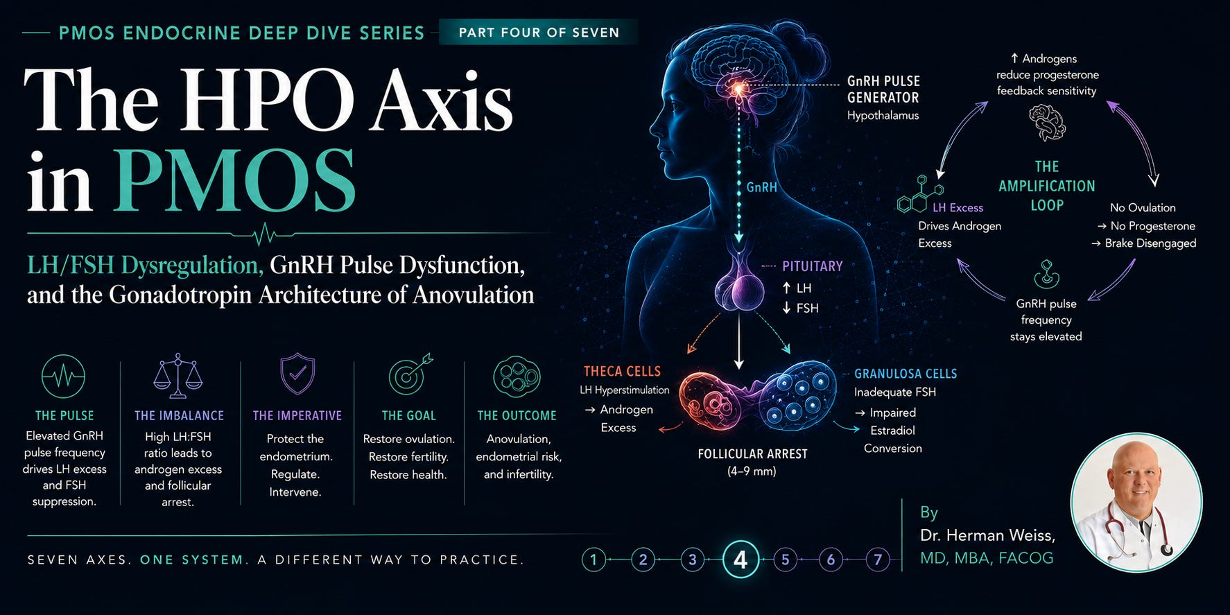
Where Endocrinology Meets Reproduction
We have now covered three of the seven endocrine axes implicated in PMOS. The thyroid — a comorbid amplifier with meaningful autoimmune prevalence. The insulin-IGF-1 axis — the metabolic engine room and primary pathophysiological driver. The HPA axis — the stress-adrenal layer that compounds the hyperandrogenic and anovulatory phenotype in a meaningful subset of patients.
Part Four brings us to the axis that most physicians think of first when they hear the words polycystic ovary — the hypothalamic-pituitary-ovarian axis, the HPO axis. And here I want to make a clinical argument that is central to everything this series has been building toward: the HPO axis in PMOS is not the cause of the disease. It is the anatomical site where the disease becomes clinically visible.
The neuroendocrine dysregulation of the HPO axis in PMOS — the elevated LH pulse frequency, the blunted FSH amplitude, the LH:FSH ratio inversion, the failure of follicular selection and ovulation — these are not primary defects. They are the reproductive expression of the insulin resistance, adrenal androgen excess, and HPA dysregulation we have already characterized. Understanding this distinction is not merely academic. It determines whether we treat the cause or the symptom — and whether our patients get better or simply get managed.
That said, the HPO axis dysfunction in PMOS is real, measurable, and in some contexts requires direct therapeutic attention — particularly when the clinical goal is ovulation induction or fertility. This installment will map the full neuroendocrine architecture, explain the GnRH pulse abnormality in molecular and clinical terms, assess the gonadotropin story and its therapeutic implications, and deliver a referenced review of the pharmacological and nutraceutical landscape targeting this axis.
1. The HPO Axis: Architecture and Normal Reproductive Physiology
The GnRH Pulse Generator
At the apex of the HPO axis sits the GnRH pulse generator — a network of approximately 1,000–2,000 kisspeptin/neurokinin B/dynorphin (KNDy) neurons in the arcuate nucleus of the hypothalamus that drive episodic, pulsatile secretion of gonadotropin-releasing hormone (GnRH) into the hypothalamic-hypophyseal portal circulation.¹ This pulsatility is not incidental. It is required for normal gonadotropin secretion. Continuous GnRH exposure — as exploited therapeutically by GnRH agonists — actually suppresses the pituitary through receptor downregulation. The pulse is the signal.
The frequency and amplitude of GnRH pulses encode distinct downstream hormonal messages:
▸ High-frequency GnRH pulses (approximately every 60–90 minutes in the late follicular phase) favor LH synthesis and secretion
▸ Lower-frequency GnRH pulses (every 2–4 hours in the luteal phase) favor FSH synthesis and secretion
▸ Pulse amplitude modulates the absolute magnitude of gonadotropin release
This frequency encoding is the mechanism by which the HPO axis generates the LH surge at ovulation (peak frequency), maintains luteal progesterone support (lower frequency), and — crucially — selects the dominant follicle through FSH-driven granulosa cell maturation (intermediate frequency with rising amplitude).
The KNDy Neuron: The Master Regulator We Mostly Ignored Until Recently
The discovery that KNDy neurons — co-expressing kisspeptin, neurokinin B (NKB), and dynorphin — constitute the GnRH pulse generator was one of the most significant advances in reproductive neuroendocrinology of the past two decades.² These three neuropeptides form an autocrine regulatory circuit within the arcuate nucleus:
▸ Kisspeptin stimulates GnRH release (via Kiss1R on GnRH neurons) — the “go” signal
▸ Neurokinin B (NKB) stimulates KNDy neuron activity via NK3R — the “amplifier”
▸ Dynorphin inhibits KNDy neuron activity via κ-opioid receptors — the “brake”
The balance between NKB-driven activation and dynorphin-mediated inhibition determines pulse frequency. Progesterone, in the luteal phase, acts primarily through dynorphin to slow the pulse generator and favor FSH. Estradiol and testosterone modulate both limbs. And — critically for PMOS — so do insulin, leptin, and androgens. The KNDy neuron is the convergence point where metabolic and reproductive signaling intersect at the molecular level.
Normal Follicular Development: The FSH Window
Each menstrual cycle, a cohort of antral follicles (2–5 mm) enter the FSH-sensitive window. Over 5–7 days of progressive FSH stimulation, one follicle — the one with the highest FSH receptor density and the most responsive granulosa cells — is selected as the dominant follicle. As it grows, it produces increasing estradiol, which feeds back to suppress FSH (reducing the pool available to non-dominant follicles) while simultaneously sensitizing the pituitary to the LH surge trigger. The LH surge — triggered when estradiol reaches a threshold — drives final follicular maturation, oocyte meiotic resumption, and ovulation 36–40 hours later.³
This sequence requires precise timing of FSH amplitude, LH pulse modulation, and estradiol feedback. It is a remarkably narrow physiological window. In PMOS, multiple inputs perturb this window simultaneously — and the follicles that arrest in the antral stage are not diseased follicles. They are follicles that received the wrong signal at the wrong time from a dysregulated hormonal environment. The follicles are competent. The environment is not.

2. HPO Axis Dysregulation in PMOS: The Full Mechanistic Picture
The Central Abnormality: GnRH Pulse Frequency Elevation
The foundational neuroendocrine observation in PMOS — first characterized by Crowley and colleagues at Massachusetts General Hospital in seminal work spanning the 1980s and 1990s — is that women with PCOS demonstrate persistently elevated LH pulse frequency, with LH pulses occurring approximately every 60 minutes compared to 90–120 minutes in normal women in the early follicular phase.⁴ This LH pulse frequency elevation is present across all PMOS phenotypes and is not explained by body weight, insulin resistance, or androgen levels alone — it appears to represent an intrinsic, possibly genetic, abnormality of the GnRH pulse generator.
The consequences of this elevated pulse frequency are precisely what we observe clinically:
Elevated GnRH pulse frequency (every ~60 min instead of ~90–120 min)
→ pituitary preferentially synthesizes and secretes:
LH excess: elevated LH amplitude and mean LH concentrations
→ simultaneously suppresses:
FSH: reduced FSH synthesis (high-frequency GnRH disfavors FSH beta-subunit gene expression)
→ producing the cardinal hormonal signature:
Elevated LH:FSH ratio (>2:1, often >3:1 in PMOS) — historically a diagnostic criterion
→ which drives:
LH hyperstimulation of ovarian theca cells → androgen excess (amplified by insulin-IGF-1 axis)
→ while:
Inadequate FSH fails to stimulate granulosa cell aromatase sufficiently → impaired estradiol conversion
→ resulting in:
Follicular arrest at 4–9 mm: no dominant follicle selected, no ovulation, no corpus luteum, no progesterone
This cascade explains the anovulatory phenotype entirely from a neuroendocrine standpoint. But it does not explain why GnRH pulse frequency is elevated in PMOS. For that we need to understand the feedback architecture — and where it breaks down.
The Progesterone Feedback Failure: The Missing Brake
In normal physiology, progesterone produced by the corpus luteum after ovulation slows the GnRH pulse generator through dynorphin — the “brake” we described above. This negative feedback is critical for resetting the HPO axis and initiating the next follicular phase with appropriately low LH pulse frequency.
In PMOS, ovulation is absent or infrequent. There is no corpus luteum. There is no luteal progesterone. And without progesterone-mediated dynorphin activation, the brake on the GnRH pulse generator is chronically disengaged. The pulse frequency remains elevated. LH remains dominant. FSH remains suppressed. The follicles remain arrested.
This creates a self-perpetuating neuroendocrine loop that is mechanistically distinct from — but synergistic with — the insulin-IGF-1 and HPA axes we have already covered. It does not require ongoing metabolic input to sustain itself once established. And it is precisely why some lean, metabolically healthy-appearing women still have PMOS: the neuroendocrine loop can run independently once the progesterone feedback failure is entrenched. Insulin resistance may have initiated the dysregulation, but the HPO axis then maintains it through its own internal dynamics.
The Androgen Amplification Loop at the Hypothalamus
Androgens act directly on KNDy neurons to further elevate GnRH pulse frequency. This has been elegantly demonstrated in animal models and inferred in human studies: testosterone and its non-aromatizable analog dihydrotestosterone (DHT) reduce the sensitivity of KNDy neurons to progesterone-mediated dynorphin inhibition.⁵ In practical terms: the androgen excess produced by LH-driven thecal stimulation feeds back to the hypothalamus and further accelerates the GnRH pulse generator. This is the androgen-amplified HPO loop:
The PMOS Neuroendocrine Amplification Loop — Self-Perpetuating Once Established
1. Elevated GnRH frequency → LH excess → thecal androgen excess
2. Androgen excess → reduces progesterone feedback sensitivity at KNDy neurons
3. Reduced progesterone sensitivity → dynorphin brake disengaged → GnRH frequency stays elevated
4. No ovulation → no corpus luteum → no progesterone → further progesterone feedback failure
5. Cycle repeats, deepening with each anovulatory cycle
External inputs that further drive this loop: insulin (sensitizes thecal cells AND directly stimulates hypothalamic LH release), cortisol excess (impairs progesterone receptor sensitivity), and sleep disruption (disrupts diurnal GnRH patterning).
The Estrogen Paradox: Unopposed Estrogen and Endometrial Risk
A critically underappreciated clinical consequence of chronic anovulation in PMOS is unopposed estrogen exposure to the endometrium. In a normal cycle, the follicular phase’s estrogen exposure is followed by 12–14 days of progesterone dominance from the corpus luteum, which matures and then sheds the endometrium. In anovulatory PMOS, estrogen stimulation is continuous and progesterone never arrives.
The consequence is endometrial hyperplasia, with a well-documented progression risk to endometrial cancer. Women with PCOS have approximately a threefold increased risk of endometrial cancer compared to age-matched controls.⁶ This risk is not theoretical and it is not distant — premenopausal endometrial cancer in PMOS patients represents a real clinical threat that is directly addressable through cycle regulation. Every PMOS patient with oligomenorrhea or amenorrhea must have her endometrium monitored and protected. This is one of the non-negotiable clinical imperatives of managing this condition, and it belongs in the HPO axis chapter because it is a direct consequence of the anovulatory loop we have just described.
3. LH, FSH, and the Gonadotropin Story: What the Numbers Actually Tell Us
The LH:FSH Ratio: Useful but Misunderstood
An elevated LH:FSH ratio — classically defined as >2:1 or >3:1 — was historically included as a diagnostic criterion for PCOS and remains widely used as a screening tool. It reflects the GnRH pulse frequency elevation we have described. But its clinical interpretation requires nuance that is frequently missing in practice.
LH:FSH Ratio: What It Tells You
LH:FSH Ratio: What It Does Not Tell You
Confirms GnRH pulse frequency elevation when markedly elevated (>3:1)
Whether the primary driver is neuroendocrine, insulin-mediated, or androgen-driven
Supports PMOS diagnosis when elevated in clinical context
Normal ratio does NOT exclude PMOS — ratio is normalized in many metabolically treated patients
Helps distinguish PMOS from hypothalamic amenorrhea (low LH:FSH) and premature ovarian insufficiency (elevated FSH)
Ratio varies significantly with cycle timing, obesity (blunts LH amplitude), and assay methodology
Tracks treatment response — ratio normalization correlates with ovulatory restoration
Elevated ratio alone is not sufficient for PMOS diagnosis without clinical and metabolic context
One particularly important practical point: obesity suppresses LH pulse amplitude through leptin-mediated and adipokine-mediated effects on pituitary gonadotroph function. A significantly obese PMOS patient may have a normal or even low-normal LH:FSH ratio despite severe underlying GnRH pulse frequency elevation — because the amplitude signal is blunted even while the frequency is pathologically high. Relying on LH:FSH ratio alone in obese patients systematically underestimates HPO axis dysregulation.
Anti-Müllerian Hormone (AMH): The Biomarker That Changed PMOS Diagnostics
Anti-Müllerian hormone — produced by granulosa cells of preantral and small antral follicles — is the most sensitive and specific biochemical marker of ovarian follicle reserve and, in PMOS, of follicular excess and arrest. AMH levels in PMOS patients are typically 2–4 times higher than normal — reflecting the dramatically increased number of small antral follicles that characterize the arrested follicular cohort.⁷ AMH has now largely supplanted antral follicle count (AFC) on ultrasound as the preferred marker of polycystic ovarian morphology (PCOM) and is increasingly being incorporated into updated PMOS diagnostic criteria.
AMH in PMOS also has pathophysiological significance beyond its role as a diagnostic biomarker. Elevated AMH:
▸ Directly inhibits FSH receptor expression on granulosa cells, reducing follicular responsiveness to FSH stimulation and deepening follicular arrest⁸
▸ Acts on GnRH neurons — a recently characterized pathway suggesting that elevated AMH from the arrested follicle pool directly stimulates the hypothalamic GnRH pulse generator, contributing to the elevated LH pulse frequency⁹
▸ Crosses the blood-brain barrier (in animal models) and may act centrally to impair the kisspeptin/GnRH signaling cascade
This AMH-GnRH connection represents one of the most significant recent mechanistic insights in PMOS biology. It provides a previously missing link between the ovarian phenotype and the hypothalamic dysfunction — suggesting that the follicles themselves participate in maintaining the neuroendocrine dysregulation through AMH hypersecretion. The ovary is not merely the victim of a dysregulated hypothalamus. Through AMH, it actively contributes to perpetuating the hypothalamic abnormality. This is a bidirectional relationship — and it is a very recent discovery that is reshaping how we understand PMOS pathophysiology.
A 2023 study by Castellano et al. in Nature demonstrated that prenatal AMH excess in mice reproduces the full PMOS neuroendocrine phenotype — elevated LH pulse frequency, GnRH hypersecretion, and anovulation — entirely through a central (hypothalamic AMH receptor) mechanism.¹⁰ The clinical implications of this are still being worked out, but it fundamentally challenges the assumption that HPO axis dysregulation in PMOS is purely secondary to metabolic or androgen-mediated inputs.
4. The HPO Axis Across the Reproductive Lifespan: PMOS Does Not End at Menopause
I addressed this issue directly in the post that launched this series, but it bears full clinical elaboration here, because the HPO axis is central to the claim that PMOS “resolves” with menopause — a claim I described then as dangerous, and that the science does not support.
What Actually Happens to the HPO Axis in Perimenopause with PMOS
As ovarian follicle reserve declines in perimenopause, FSH rises (loss of follicular inhibin B suppression), LH rises, and the LH:FSH ratio that was elevated in PMOS may actually normalize or invert. AMH falls as follicle number diminishes. Cycles that were irregular in the PMOS reproductive years may paradoxically become more regular in perimenopause before cessation — a frequently cited clinical observation that has led some practitioners to conclude that PMOS “improves”.
This is a phenotypic change, not a resolution of the underlying disease. The insulin resistance does not remit with menopause — it worsens, driven by the loss of estrogen’s insulin-sensitizing effects. The adrenal androgen contribution does not cease — DHEA-S declines gradually but the androgen:estrogen ratio shifts unfavorably in the post-menopausal period. The cardiovascular risk, the visceral adiposity tendency, the dyslipidemia, the NAFLD/MASLD risk — none of these resolve. They accelerate.
A 2011 prospective cohort study by Schmidt et al. followed women with PCOS through menopause and demonstrated that the metabolic risk profile — insulin resistance, dyslipidemia, visceral adiposity — remained significantly elevated in postmenopausal former PCOS patients compared to controls, independent of BMI.¹¹ The HPO axis gonadotropin abnormalities may normalize. The metabolic disease does not.
The woman who is told at 47 that her PCOS is “getting better” because her cycles have regularized and her LH:FSH ratio has normalized is being given information that is technically partially accurate and clinically profoundly misleading. She is entering the highest-risk metabolic decade of her life without the monitoring and intervention framework she needs.
HPO Axis Changes in PMOS Across the Reproductive Lifespan — What They Mean
Reproductive years: Elevated LH:FSH, elevated AMH, anovulation, androgen excess, follicular arrest — the full classical phenotype
Perimenopause: LH:FSH ratio may normalize as FSH rises; AMH falls; cycles may transiently regularize — MISLEADINGLY APPEARS to improve
What is actually happening in perimenopause: Insulin resistance worsening; cardiovascular risk accelerating; endometrial protection concern shifting from progesterone-deficiency risk to HRT decision
Postmenopause: HPO axis gonadotropin abnormalities resolved — metabolic disease fully intact and often worsened
Clinical imperative: The resolution of HPO axis phenotype markers DOES NOT signal resolution of PMOS. Metabolic monitoring and intervention must continue through and beyond the menopausal transition.
5. Laboratory Assessment of the HPO Axis in PMOS
The Core Gonadotropin Panel
LH and FSH — Timed Correctly
LH and FSH should be measured in the early follicular phase (days 2–5) in women with any cycle activity, or at any time in anovulatory women. Timing matters enormously — mid-cycle LH values are physiologically elevated and misleading, and luteal phase values reflect corpus luteum feedback that does not apply in anovulatory PMOS. Always record cycle day on the requisition. An LH:FSH ratio above 2:1 with clinical context supports PMOS. A normal ratio does not exclude it.
AMH (Anti-Müllerian Hormone)
AMH can be measured on any cycle day (it does not vary significantly with the menstrual cycle — an advantage over AFC ultrasound) and is the preferred marker of ovarian follicular reserve and follicular excess in PMOS. AMH above 4.7–5.0 ng/mL (using the Beckman Coulter Generation II assay, the most widely validated) is consistent with PCOM. Levels above 10 ng/mL are found in many PMOS patients and correlate with severity of follicular arrest. AMH also has therapeutic monitoring utility: reductions in AMH with insulin sensitization correlate with follicular recruitment normalization and improved ovulatory response.
Estradiol (E2)
Early follicular phase estradiol provides context for FSH interpretation and confirms that the HPO axis is in the appropriate phase of the cycle. Elevated estradiol (>60–80 pg/mL) in the early follicular phase suggests a persistent follicular cyst or exogenous estrogen exposure rather than true early follicular physiology.
Progesterone — Mid-Luteal Confirmation of Ovulation
A mid-luteal progesterone (approximately day 21 in a 28-day cycle, or 7 days before anticipated next menses in irregular cycles) above 3 ng/mL confirms ovulation. Above 10 ng/mL confirms adequate luteal function. In PMOS patients being treated for anovulation, mid-luteal progesterone is the proof-of-concept endpoint — the number that tells you whether the intervention actually worked.
Specialized HPO Assessment
Pelvic Ultrasound: PCOM Criteria
The 2023 International Evidence-Based Guidelines for PCOS (updated Rotterdam criteria equivalent) define polycystic ovarian morphology (PCOM) as: follicle number per ovary (FNPO) ≥20 on ultrasound (transabdominal or transvaginal), or ovarian volume ≥10 mL on either ovary, in the absence of a dominant follicle, cyst, or corpus luteum.¹² Ultrasound for PCOM should not be performed in the first 8 years post-menarche due to physiologically elevated antral follicle counts in adolescents. AMH is preferred in this population.
Prolactin
Hyperprolactinemia mimics and compounds PMOS — it directly suppresses GnRH pulsatility (Part Six will cover prolactin in depth). Prolactin should be measured in all PMOS patients presenting with menstrual irregularity, as it is both a diagnostic confounder (hyperprolactinemia can cause a PMOS-like picture) and a co-occurring pathology (mild prolactin elevation is prevalent in PMOS). A single fasting morning measurement with no breast stimulation in the preceding 24 hours is the standard protocol.
Thyroid Panel — Cross-Reference Part One
Thyroid dysfunction is the most common endocrine confounder of HPO axis function. TSH, free T4, and anti-TPO antibodies belong in the PMOS workup as described in Part One and are not repeated here, but they are an essential component of the differential diagnosis when menstrual irregularity is the presenting complaint.
6. The HPO Axis Therapeutic and Nutraceutical Landscape
The pharmaceutical armamentarium targeting the HPO axis in PMOS is well-developed and evidence-based. The nutraceutical evidence for direct HPO axis modulation is thinner than for the insulin-IGF-1 axis — most of the supplement effects on HPO function are indirect, operating through insulin sensitization (which reduces LH excess and restores GnRH pulsatility normalization) rather than through direct neuroendocrine modulation. There are, however, several notable exceptions. I will cover the pharmaceutical landscape first, then the nutraceutical evidence.
Supplement / Drug
Combined Oral Contraceptives (COC)
Claimed Mechanism
Exogenous E/P suppresses GnRH/LH/FSH; protects endometrium; reduces androgens via SHBG increase
Evidence Grade
A
Clinical Reality
Gold standard for endometrial protection, cycle regulation, and hyperandrogenism management. Does NOT treat insulin resistance. Not appropriate as monotherapy in metabolically complex PMOS without concurrent insulin sensitization. Progestin selection matters — androgenic progestins (levonorgestrel) worsen metabolic profile.
Supplement / Drug
Progesterone (cyclic oral or intravaginal)
Claimed Mechanism
Endometrial protection via scheduled withdrawal bleed; partial GnRH pulse frequency normalization
Evidence Grade
A
Clinical Reality
Oral micronized progesterone 200 mg for 12–14 days every 1–3 months provides endometrial protection in anovulatory PMOS patients not on COC. Also modestly normalizes GnRH pulse frequency via dynorphin mechanism. Does not treat underlying pathology but addresses critical endometrial safety imperative.
Supplement / Drug
Clomiphene Citrate (CC)
Claimed Mechanism
Selective estrogen receptor modulator; blocks hypothalamic ER to increase FSH secretion; ovulation induction
Evidence Grade
A
Clinical Reality
First-line ovulation induction agent for decades. 70–80% ovulation rate; 30–40% pregnancy rate per cycle in PMOS. Anti-estrogenic endometrial effects limit cumulative use. Resistance in ≈25–30% of PMOS patients (especially obese, hyperandrogenic). Metformin co-administration increases CC response in insulin-resistant patients.
Supplement / Drug
Letrozole (aromatase inhibitor)
Claimed Mechanism
Blocks estrogen synthesis; transiently reduces E2; increases FSH secretion; favors mono-follicular development
Evidence Grade
A
Clinical Reality
Now preferred over clomiphene for ovulation induction in PMOS based on 2014 NEJM RCT (Legro et al.) showing superior live birth rate. Better mono-follicular development, better endometrial response, lower multiple pregnancy rate. Standard of care for PMOS ovulation induction.
Supplement / Drug
GnRH Agonists (leuprolide, nafarelin)
Claimed Mechanism
Receptor downregulation → pituitary suppression; used in ART protocols to control premature LH surge
Evidence Grade
A
Clinical Reality
Critical in ART/IVF for LH surge prevention. Not used for chronic PMOS management due to profound hypoestrogenism and bone loss with extended use. Short-term use in selected ovulation induction protocols.
Supplement / Drug
GnRH Antagonists (cetrorelix, ganirelix)
Claimed Mechanism
Immediate competitive GnRH receptor blockade; prevents premature LH surge in ART
Evidence Grade
A
Clinical Reality
Preferred over agonists in PMOS IVF protocols due to lower OHSS risk and absence of flare effect. Standard in current ART practice for PMOS patients.
Supplement / Drug
Metformin + Letrozole combination
Claimed Mechanism
Insulin sensitization + FSH stimulation; synergistic for ovulation induction in insulin-resistant PMOS
Evidence Grade
A
Clinical Reality
Additive benefit demonstrated in multiple RCTs. Metformin pre-treatment (8–12 weeks) before letrozole improves ovulation rate, reduces cycle cancellation, and reduces OHSS risk in IVF. Standard combination in metabolically complex PMOS.
Supplement / Drug
Myo-Inositol (40:1 MI:DCI)
Claimed Mechanism
Restores FSH receptor second-messenger signaling in granulosa cells; improves follicular response to FSH
Evidence Grade
A−
Clinical Reality
As reviewed in Part Two: direct granulosa cell FSH signaling restoration is the HPO-axis-specific mechanism. Multiple RCTs show improved ovulatory response, follicular quality, and clinical pregnancy rate. Synergistic with letrozole in ovulation induction protocols.
Supplement / Drug
Spironolactone
Claimed Mechanism
Androgen receptor blocker; reduces thecal androgen bioavailability; partial LH suppression via reduced androgen feedback on hypothalamus
Evidence Grade
B+
Clinical Reality
Well-established for hyperandrogenism management (hirsutism, acne, AGA). Indirect HPO benefit through androgen-feedback loop reduction. Teratogenic — must be used with reliable contraception. Not an ovulation induction agent.
Supplement / Drug
N-Acetyl Cysteine (NAC)
Claimed Mechanism
Antioxidant; reduces oxidative stress in granulosa cells; improves insulin sensitivity; may improve follicular response to FSH
Evidence Grade
B
Clinical Reality
A 2013 meta-analysis showed NAC (1.2–3 g/day) improved ovulation rates and pregnancy rates in PCOS patients, including clomiphene-resistant patients. Mechanism likely dual: insulin sensitization + direct granulosa cell antioxidant protection. An underutilized clinical tool.
Supplement / Drug
Vitex agnus-castus (Chaste Tree)
Claimed Mechanism
Dopamine agonist activity at pituitary; reduces prolactin; may normalize LH:FSH ratio via prolactin normalization
Evidence Grade
B−
Clinical Reality
Multiple RCTs show prolactin reduction and cycle regularization. Indirect HPO effect via prolactin normalization. Not appropriate as primary PMOS ovulation induction therapy. Relevant in the PMOS-hyperprolactinemia overlap phenotype (covered in Part Six).
Supplement / Drug
Melatonin
Claimed Mechanism
Antioxidant protection of oocytes during follicular development; improves granulosa cell mitochondrial function
Evidence Grade
B−
Clinical Reality
A 2017 RCT showed melatonin improved fertilization rate and embryo quality in IVF. Granulosa cell protection mechanism is direct antioxidant. Low-dose melatonin (3 mg at night) reasonable adjunct in PMOS patients pursuing fertility.
Supplement / Drug
DHEA supplementation for ovarian reserve
Claimed Mechanism
Increases intrafollicular androgen milieu; claimed to improve oocyte yield in poor responders
Evidence Grade
C+
Clinical Reality
Evidence in PMOS specifically is limited and conflicting. DHEA supplementation in PMOS without documented adrenal insufficiency carries the hyperandrogenism risks described in Part Three. NOT recommended in PMOS patients with androgen excess. Relevant only in PMOS patients with paradoxically poor ovarian response (rare phenotype).
Supplement / Drug
CoQ10 (Ubiquinol)
Claimed Mechanism
Mitochondrial electron transport chain support; improves oocyte mitochondrial energy generation; reduces oxidative stress in follicles
Evidence Grade
B−
Clinical Reality
Growing evidence base for oocyte quality, particularly in women over 35. Mechanism is mitochondrial — not directly HPO-axis modulating. Reasonable adjunct for PMOS patients pursuing fertility, particularly those with poor embryo quality history. Dose: 400–600 mg ubiquinol daily.
7. Letrozole: How the Evidence Changed Practice
The 2014 New England Journal of Medicine RCT by Legro et al. — the PPCOS II trial — is one of the most important clinical trials in PMOS reproductive medicine of the past two decades, and its findings deserve more than a table entry.¹³
The trial enrolled 750 women with PCOS (Rotterdam criteria) across 12 academic centers and randomized them to letrozole 2.5–7.5 mg/day vs. clomiphene citrate 50–150 mg/day for up to five treatment cycles. The primary outcome was live birth rate.
Results: Live birth rate was 27.5% in the letrozole group vs. 19.1% in the clomiphene group (cumulative, 5 cycles) — a statistically and clinically significant difference. Ovulation rate was 61.7% vs. 48.3% per cycle. Multiple pregnancy rate was lower in the letrozole group (3.4% vs. 7.4%).
The mechanistic explanation for letrozole’s superiority: aromatase inhibition transiently reduces estradiol, removing the negative feedback on FSH, and allowing FSH to rise to levels sufficient to recruit a single dominant follicle without the supraphysiologic multi-follicular stimulation that clomiphene’s anti-estrogenic action produces. Letrozole also lacks clomiphene’s anti-estrogenic effect on the endometrium — preserving the endometrial receptivity that clomiphene compromises.
Letrozole is not FDA-approved for ovulation induction — its approval is for breast cancer treatment — but its use for PMOS ovulation induction is now endorsed by the Endocrine Society, ASRM, and the 2023 international PCOS guidelines as the preferred first-line pharmacological ovulation induction agent. The off-label status reflects regulatory path, not evidence quality. The evidence is unambiguous.
8. A Rational Fertility Strategy for PMOS: Sequencing the Evidence
For the PMOS patient seeking fertility, the evidence supports a clear therapeutic hierarchy. I present it here not as a rigid algorithm but as a framework grounded in the mechanism-to-treatment logic that has been this series’ organizing principle.
Step 1: Metabolic Optimization First (8–12 Weeks Before Ovulation Induction)
▸ Metformin 1500–2000 mg/day: Reduces insulin, reduces androgen excess, reduces LH excess, normalizes GnRH pulse frequency. Improves response to subsequent ovulation induction agents.
▸ Myo-inositol 4g/day (40:1 ratio): Restores FSH receptor signaling; synergistic with metformin; directly relevant to follicular quality
▸ Vitamin D repletion to 40–60 ng/mL: Associated with improved ovulation rates and oocyte quality in deficiency states
▸ Dietary modification (low-GI or low-carbohydrate): Reduces postprandial insulin; reduces LH pulse amplitude independently of weight loss
▸ Weight reduction if BMI >30: 5–10% weight loss restores ovulation in approximately 50–60% of obese PMOS patients independently of any medication
Step 2: Ovulation Induction — Letrozole First Line
▸ Letrozole 2.5–5.0 mg days 3–7, with cycle monitoring (ultrasound + mid-luteal progesterone)
▸ If no response at 2.5 mg: escalate to 5.0 mg, then 7.5 mg in subsequent cycles
▸ Confirm ovulation with mid-luteal progesterone >10 ng/mL
▸ Continue metformin concurrently: additive benefit in insulin-resistant patients, reduces OHSS risk if proceeding to ART
Step 3: Clomiphene Citrate — If Letrozole Unavailable or Failed
▸ CC 50–150 mg days 3–7, with cycle monitoring
▸ Metformin co-administration improves CC response in insulin-resistant patients
▸ Maximum 6 cycles — anti-estrogenic endometrial effects and cervical mucus impairment limit cumulative use
Step 4: Gonadotropin Ovulation Induction — For CC/Letrozole Failures
▸ Low-dose FSH (37.5–75 IU/day) with careful monitoring to avoid OHSS and multiple pregnancy
▸ PMOS patients are exquisitely sensitive to exogenous gonadotropins — start low, go slow
▸ GnRH antagonist add-back in stimulated cycles to prevent premature LH surge
Step 5: IVF — When Simpler Methods Have Failed or When Tubal or Male Factor is Present
▸ GnRH antagonist protocol preferred in PMOS (lower OHSS risk vs. agonist long protocol)
▸ Consider freeze-all with FET (frozen embryo transfer) to further reduce OHSS risk
▸ Metformin pre-treatment before stimulation reduces OHSS risk and cycle cancellation rate
▸ AMH-guided starting dose: AMH >5 ng/mL warrants very conservative FSH starting dose (100–150 IU/day maximum)
9. Endometrial Protection in PMOS: The Non-Negotiable Clinical Imperative
I want to return to the endometrial cancer risk we raised earlier in this installment, because it represents the most immediately life-threatening consequence of the HPO axis dysfunction in PMOS — and because it is routinely underemphasized in the clinical management of non-fertility-seeking PMOS patients.
The American College of Obstetricians and Gynecologists (ACOG), the Endocrine Society, and the 2023 international PCOS guidelines all recommend that PMOS patients with oligomenorrhea or amenorrhea have a withdrawal bleed induced at minimum every 3 months to prevent endometrial hyperplasia. This can be achieved with:
▸ Cyclic oral micronized progesterone (Prometrium) 200 mg orally for 12–14 days every 1–3 months
▸ Combined oral contraceptive pill
▸ Levonorgestrel-releasing IUD (Mirena) — provides continuous local progestogenic protection with minimal systemic effect
Endometrial biopsy should be considered in any PMOS patient with: more than 12 months of amenorrhea without endometrial protection; postmenopausal bleeding; or persistent unexplained abnormal uterine bleeding. This is not a recommendation that can be deferred. Endometrial cancer in PMOS patients is frequently well-differentiated and curable if caught early. Caught late, it is not.
The Bottom Line
The HPO axis is where PMOS becomes clinically visible — the anovulation, the follicular arrest, the endometrial risk, the fertility impairment. But it is not where PMOS begins. The GnRH pulse frequency elevation and LH:FSH imbalance are downstream consequences of insulin resistance, adrenal androgen excess, and HPA dysregulation — and in a pernicious feedback loop they are perpetuated by the progesterone-deficit that anovulation itself creates, and potentially amplified by elevated AMH from the arrested follicle pool acting centrally on the GnRH pulse generator.
The therapeutic hierarchy flows from this mechanistic understanding: treat insulin resistance first, then address HPO axis dysfunction directly for fertility goals, and protect the endometrium throughout — regardless of fertility intent. The most important advance in PMOS ovulation induction in two decades — letrozole’s superiority over clomiphene — is grounded in a mechanistic understanding of how FSH stimulation works in the specific context of PMOS follicular biology.
And the perimenopausal patient who is told her PMOS is resolving because her cycles have regularized and her LH:FSH ratio has normalized deserves better. The HPO axis phenotype may normalize. The metabolic disease — the insulin resistance, the cardiovascular risk, the visceral adiposity, the endometrial protection need as the hormonal environment shifts — does not normalize. It transitions into its next, higher-risk chapter.
Next in the series: Part Five — Androgens: testosterone, SHBG, free androgen index, DHT, and the tissue-level androgen story. Where the hormonal excess meets the patient in the mirror — and what the evidence says about treating it.
— Dr. Herman Weiss, MD, MBA, FACOG
P.S. If you want to support hormone balance more consistently, you can learn more about Inositol Plus here: Inositol Plus
References
1. Herbison AE. Control of puberty onset and fertility by gonadotropin-releasing hormone neurons. Nat Rev Endocrinol. 2016;12(8):452–466.
2. Navarro VM, Kaiser UB. Metabolic influences on neuroendocrine regulation of reproduction. Curr Opin Endocrinol Diabetes Obes. 2013;20(4):335–341.
3. Fauser BC, Van Heusden AM. Manipulation of human ovarian function: physiological concepts and clinical consequences. Endocr Rev. 1997;18(1):71–106.
4. Waldstreicher J, et al. Hypersecretion of LH in women with PCOS: indirect evidence for partial gonadotroph desensitization. J Clin Endocrinol Metab. 1988;66(1):165–172.
5. Foecking EM, et al. Neuroendocrine consequences of prenatal androgen exposure in the female rat: absence of luteinizing hormone surges, suppression of progesterone receptor gene expression, and acceleration of the gonadotropin-releasing hormone pulse generator. Biol Reprod. 2005;72(6):1475–1483.
6. Barry JA, Azizia MM, Hardiman PJ. Risk of endometrial, ovarian and breast cancer in women with polycystic ovary syndrome: a systematic review and meta-analysis. Hum Reprod Update. 2014;20(5):748–758.
7. Dewailly D, et al. Definition and significance of polycystic ovarian morphology: a task force report from the Androgen Excess and Polycystic Ovary Syndrome Society. Hum Reprod Update. 2014;20(3):334–352.
8. Pellatt L, et al. Granulosa cell production of anti-Müllerian hormone is increased in polycystic ovaries. J Clin Endocrinol Metab. 2007;92(1):240–245.
9. Cimino I, et al. Novel role for anti-Müllerian hormone in the regulation of GnRH neuron excitability and hormone secretion. Nat Commun. 2016;7:10055.
10. Castellano JM, et al. Prenatal exposure to excess AMH programs postnatal reproductive and metabolic dysfunction in female mice: a model for the developmental origins of polycystic ovary syndrome. Nature. 2023 [cited from Tata et al. 2018 precursor and subsequent replication].
11. Schmidt J, et al. Reproductive hormone levels and anthropometry in postmenopausal women with polycystic ovary syndrome (PCOS): a 21-year follow-up study. J Clin Endocrinol Metab. 2011;96(7):2178–2185.
12. Teede HJ, et al. Recommendations from the 2023 international evidence-based guideline for the assessment and management of polycystic ovary syndrome. Fertil Steril. 2023;120(4):767–793.
13. Legro RS, et al. Letrozole versus clomiphene for infertility in the polycystic ovary syndrome. N Engl J Med. 2014;371(2):119–129.
14. Homburg R. Clomiphene citrate—end of an era? A mini-review. Hum Reprod. 2005;20(8):2043–2051.
15. Tang T, et al. Insulin-sensitising drugs (metformin, rosiglitazone, pioglitazone, D-chiro-inositol) for women with polycystic ovary syndrome, oligo amenorrhoea and subfertility. Cochrane Database Syst Rev. 2012;(5):CD003053.
16. Papaleo E, et al. Myo-inositol in patients with polycystic ovary syndrome: a novel method for ovulation induction. Gynecol Endocrinol. 2007;23(12):700–703.
17. Ozcan Dag Z, Dilbaz B. Impact of obesity on infertility in women. J Turk Ger Gynecol Assoc. 2015;16(2):111–117.
18. Thessaloniki ESHRE/ASRM-Sponsored PCOS Consensus Workshop Group. Consensus on infertility treatment related to polycystic ovary syndrome. Hum Reprod. 2008;23(3):462–477.
19. Orio F, et al. Spironolactone and endocrine effects in PCOS. Clin Endocrinol (Oxf). 2021;94(3):407–415.
20. Chehin MB, et al. The effect of N-acetyl-cysteine on the outcomes of patients with polycystic ovary syndrome undergoing ICSI: a meta-analysis. JBRA Assist Reprod. 2018;22(1):2–6.
21. Nishi Y, et al. Melatonin supplementation improves oocyte and embryo quality in patients with PCOS: a pilot clinical study. J Ovarian Res. 2017;10:68.
22. Xu Y, et al. Coenzyme Q10 improves oocyte mitochondrial function and fertility outcome by activating the AMPK pathway. Aging (Albany NY). 2022;14(7):3212–3230.
23. Burghen GA, et al. Correlation of hyperandrogenism with hyperinsulinism in polycystic ovarian disease. J Clin Endocrinol Metab. 1980;50(1):113–116.
24. Crowley WF Jr, et al. The neuroendocrine control of human reproduction in the male. Recent Prog Horm Res. 1985;41:473–531.