THE HPA AXIS IN PMOS
Cortisol, Adrenal Androgens, and DHEA-S: Why Stress Physiology Is a Biochemical Problem, Not a Psychological One

The Axis Nobody Wants to Talk About
In Part Two, we established that the insulin-IGF-1 axis is the engine room of PMOS, the central pathophysiological driver from which hyperandrogenism, anovulation, and metabolic dysfunction all cascade. In Part One, we addressed the thyroid: a meaningful amplifier, a genuine comorbidity, frequently under-assessed.
Now we arrive at the hypothalamic-pituitary-adrenal axis, the HPA axis, and I want to begin by naming something directly: this is the axis most frequently co-opted by wellness culture, the supplement industry, and the “adrenal fatigue” ecosystem into a framework that is simultaneously partially correct and substantively distorted. Partially correct, because the HPA axis is genuinely dysregulated in a meaningful proportion of PMOS patients, and that dysregulation has real clinical consequences. Substantively distorted, because the framing of cortisol dysfunction in wellness culture almost invariably outpaces what the evidence actually supports.
My commitment in this series has been to read the evidence straight, neither dismiss what is real nor inflate what is preliminary. Nowhere in this series does that commitment require more discipline than here. So let us be precise.
1. The HPA Axis: Architecture and Normal Physiology
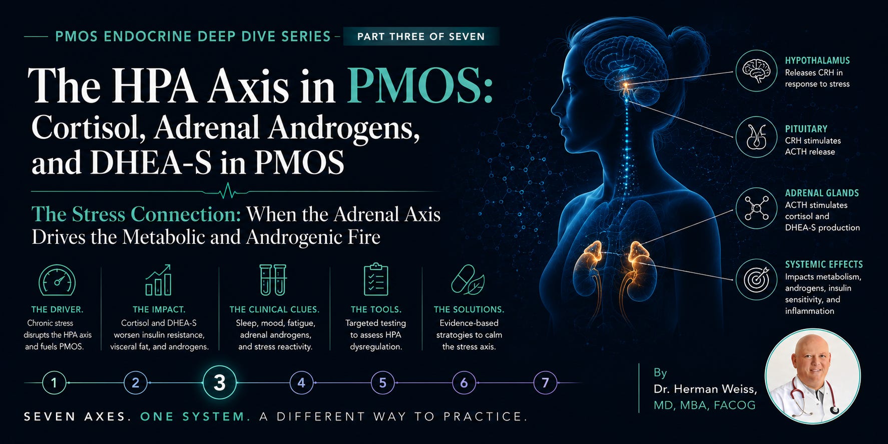
The hypothalamic-pituitary-adrenal axis is a neuroendocrine cascade governing the stress response, energy metabolism, immune modulation, and reproductive function. Its architecture is straightforward:
Hypothalamus: secretes Corticotropin-Releasing Hormone (CRH) in response to stress, circadian cues, cytokines, and glucose signals
→ stimulates:
Anterior Pituitary: releases Adrenocorticotropic Hormone (ACTH)
→ stimulates:
Adrenal Cortex (Zona Fasciculata): produces cortisol, the primary glucocorticoid
Adrenal Cortex (Zona Reticularis): produces adrenal androgens — DHEA, DHEA-S, androstenedione
→ cortisol feeds back to:
Hypothalamus and Pituitary: negative feedback inhibiting further CRH and ACTH secretion (the long-loop feedback)
Normal cortisol follows a diurnal rhythm: peak at 6–8 AM (the cortisol awakening response, or CAR), gradual decline through the day, nadir around midnight. This rhythm is not merely about energy — it is a master regulator of metabolic, immune, and reproductive function. Disruption of this rhythm — whether from chronic psychosocial stress, sleep dysregulation, or intrinsic HPA axis dysfunction — has downstream consequences that are mechanistically traceable and clinically significant.
The Adrenal Androgen Pathway: DHEA, DHEA-S, and Androstenedione
The adrenal zona reticularis is an often-overlooked androgen factory. Under ACTH stimulation, it produces dehydroepiandrosterone (DHEA) and its sulfated form DHEA-S, plus androstenedione, all weak androgens that serve as precursors to testosterone and estradiol in peripheral tissues. In reproductive-age women, the adrenal gland contributes approximately 50% of circulating DHEA-S and a significant fraction of androstenedione.¹
This matters profoundly in PMOS. When we identify hyperandrogenism in a PMOS patient, we cannot assume the ovary is the sole source. In approximately 20–35% of PMOS patients, adrenal androgen excess, sometimes called adrenal PMOS or the adrenal PMOS phenotype, is the primary or a co-primary driver of the hyperandrogenic state.² This distinction is not academic. It has direct implications for which treatments are most likely to work.

2. HPA Axis Dysregulation in PMOS: The Evidence
Three distinct but interacting patterns of HPA dysregulation have been documented in PMOS, each with its own mechanistic basis and clinical signature. Understanding which pattern predominates in a given patient is the key to rational therapeutic decision-making.
Pattern 1: CRH Hypersensitivity and Adrenocortical Hyperresponsiveness
The most consistently documented HPA abnormality in PMOS is an exaggerated adrenocortical response to ACTH stimulation, specifically, elevated DHEA-S and androstenedione responses to both exogenous ACTH challenge and to CRH stimulation tests, in the context of normal or only modestly elevated basal cortisol.³
Azziz et al. demonstrated in a foundational study that women with PCOS with elevated DHEA-S showed exaggerated adrenal androgen responses to ACTH stimulation, disproportionate to the cortisol response, suggesting selective adrenal androgen hyperresponsiveness rather than global HPA axis overactivation.⁴ The molecular basis for this appears to involve dysregulation of CYP11A1 and CYP17A1 in the adrenal cortex, the same cytochrome P450 enzymes implicated in ovarian androgen excess, suggesting a shared enzymatic vulnerability across adrenal and ovarian steroidogenesis in PMOS.
This is mechanistically important: it means the same serine kinase dysregulation identified in insulin receptor signaling and ovarian CYP17A1 hyperactivity (as described in Part Two) may also operate in adrenal steroidogenesis. PMOS may involve a shared enzymatic abnormality across multiple steroidogenic tissues — one metabolic defect, multiple phenotypic expressions. This is the polyendocrine story told at the molecular level.
Pattern 2: Hypercortisolism, Visceral Adiposity, and the Metabolic Feedforward
A subset of PMOS patients — particularly those with obesity, significant visceral adiposity, and severe insulin resistance — show evidence of mild to moderate hypercortisolism, often without meeting criteria for Cushing’s syndrome. This is not Cushing’s disease. But it is not nothing.
Pasquali et al. documented elevated 24-hour urinary free cortisol and exaggerated cortisol responses to CRH in obese PCOS patients compared to weight-matched controls without PCOS.⁵ The proposed mechanism involves enhanced cortisol regeneration in visceral adipose tissue, mediated by the enzyme 11β-hydroxysteroid dehydrogenase type 1 (11β-HSD1), which converts inactive cortisone to active cortisol. Visceral fat expresses high levels of 11β-HSD1 — creating a local hypercortisolaemic microenvironment that amplifies insulin resistance, promotes further visceral fat deposition, and stimulates adrenal androgen production.⁶
This is a critically underappreciated feedforward loop in PMOS: visceral fat → local cortisol regeneration via 11β-HSD1 → increased insulin resistance → more visceral fat → more 11β-HSD1 activity. The clinical consequence:
▸ A PMOS patient with significant visceral adiposity may have functionally elevated tissue cortisol even with a normal serum cortisol measurement
▸ Weight loss in this patient reduces 11β-HSD1 activity, reduces local cortisol exposure, improves insulin sensitivity, and reduces adrenal androgen output — simultaneously and through a single mechanism
▸ 11β-HSD1 inhibition is an active drug development target for metabolic syndrome and PMOS for precisely this reason
Pattern 3: Dysregulated Diurnal Rhythm and the Sleep-Cortisol-Insulin Triangle
The third pattern is the one most commonly encountered in clinical practice and the one most directly addressable by non-pharmacological means: disruption of the normal cortisol diurnal rhythm, most commonly manifest as blunted cortisol awakening response (CAR), elevated evening cortisol, or both. This pattern is closely linked to sleep dysregulation — which is itself highly prevalent in PMOS.
Obstructive sleep apnea (OSA) affects 50–70% of obese women with PCOS and a clinically significant proportion of lean patients — a rate far exceeding the general female population matched for BMI and age.⁷ OSA drives nocturnal cortisol spikes through hypoxia-triggered HPA activation, disrupts growth hormone pulsatility, worsens insulin resistance through sleep fragmentation, and impairs glucose metabolism through multiple simultaneous mechanisms. A PMOS patient with undiagnosed OSA is fighting multiple metabolic battles simultaneously, several of which operate through cortisol.
Beyond OSA, chronic psychosocial stress — whether from the PMOS diagnosis itself, its symptomatic burden (weight gain, infertility, hirsutism, acne), or external life stressors — maintains HPA axis activation at low but chronically elevated levels. Chronic low-grade hypercortisolaemia of this kind does not typically produce the dramatic phenotype of Cushing’s. What it produces is a metabolic environment characterized by: increased hepatic gluconeogenesis, reduced peripheral insulin sensitivity, increased appetite and preferential caloric deposition in visceral fat, suppressed reproductive axis activity, and reduced immune regulatory capacity. These are not soft findings. They are well-characterized glucocorticoid pharmacology operating at endogenous cortisol levels.
The Three HPA Dysregulation Patterns in PMOS — At a Glance
Pattern 1 — Adrenal Androgen Hyperresponsiveness: Elevated DHEA-S and androstenedione response to ACTH; normal or modestly elevated basal cortisol; shared CYP17A1 dysregulation with ovarian steroidogenesis. (~20–35% of PMOS patients)
Pattern 2 — 11β-HSD1-Mediated Visceral Hypercortisolism: Elevated tissue cortisol regeneration in visceral fat; amplifies insulin resistance and adrenal androgens; may show normal serum cortisol despite significant local cortisol excess. (Predominates in obese/high-visceral-fat phenotype)
Pattern 3 — Dysregulated Diurnal Rhythm: Blunted cortisol awakening response; elevated evening cortisol; closely linked to OSA, sleep dysregulation, and chronic psychosocial stress. (Prevalent across PMOS phenotypes; most clinically addressable)
3. Cortisol and the Reproductive Axis: The Direct Suppression Pathway
The HPA-HPO interface is not a peripheral concern in PMOS. It is a direct mechanistic connection with documented pathways at every level of the reproductive axis.
At the Hypothalamic Level
CRH directly inhibits GnRH pulsatility — a well-characterized mechanism in stress-induced hypothalamic amenorrhea. In PMOS, the context is more complex: baseline GnRH pulse frequency is already elevated (driving the LH:FSH imbalance described in Part Two), but chronic CRH excess can paradoxically both stimulate androgen-driven LH excess and suppress the FSH amplitude needed for follicular selection, depending on the pattern and timing of HPA activation.⁸
At the Pituitary Level
Glucocorticoids exert direct inhibitory effects on pituitary LH and FSH secretion through glucocorticoid receptor-mediated transcriptional suppression of gonadotropin gene expression.⁹ Chronic hypercortisolaemia therefore blunts the gonadotropin amplitude needed for follicular maturation and ovulation — adding a pituitary-level contribution to the anovulatory phenotype on top of the hypothalamic GnRH abnormality.
At the Ovarian Level
The ovary expresses glucocorticoid receptors. Direct glucocorticoid action on ovarian granulosa and thecal cells modulates steroidogenesis and follicular survival. Elevated cortisol at the ovarian level impairs progesterone synthesis in granulosa cells and reduces oocyte quality through glucocorticoid-receptor-mediated apoptosis signaling.¹⁰
This means that in a PMOS patient with significant HPA dysregulation, cortisol is acting simultaneously at the hypothalamus, the pituitary, and the ovary to compound the anovulatory phenotype that insulin-IGF-1 dysregulation initiated. The ovarian dysfunction in PMOS is therefore not a two-input system (insulin + LH). In a meaningful subset of patients, it is a three-input system: insulin + LH imbalance + cortisol. Treating insulin resistance alone in the high-HPA-dysregulation phenotype may produce incomplete clinical response. This is not a treatment failure. It is a diagnosis failure — we missed an active contributing axis.
4. DHEA-S in PMOS: The Most Misunderstood Androgen
DHEA-S is the most abundant circulating steroid in the human body and the primary indicator of adrenal androgen activity. It is produced almost exclusively by the adrenal zona reticularis, has a long half-life (8–10 hours, compared to hours for DHEA), and serves as a reservoir for peripheral androgen conversion to testosterone and estradiol.
In PMOS, elevated DHEA-S is found in approximately 20–30% of patients and is a cardinal marker of adrenal androgen excess, distinguishing the adrenal PMOS phenotype from the predominantly ovarian phenotype.¹¹ Yet DHEA-S is simultaneously one of the most misinterpreted lab values in women’s health — both under-assessed in conventional gynecology and radically over-supplemented in the wellness and anti-aging space.
The DHEA-S Testing Imperative
DHEA-S should be measured in every PMOS patient as part of the androgen panel. This is not standard practice in all settings, and it should be. An elevated DHEA-S (above 350–400 mcg/dL in reproductive-age women, though laboratory reference ranges vary) signals adrenal androgen excess and should prompt consideration of: adrenal androgen suppression therapy; exclusion of non-classical congenital adrenal hyperplasia (ncCAH) via 17-hydroxyprogesterone measurement; and screening for cortisol axis abnormalities. It also changes the therapeutic hierarchy — a patient whose hyperandrogenism is primarily adrenal-driven will not respond optimally to ovarian suppression alone.
The DHEA Supplementation Problem
DHEA is sold over-the-counter in the United States as a dietary supplement — a regulatory anomaly that does not exist in most other developed countries, where DHEA is appropriately classified as a prescription hormone. The supplement market for DHEA is enormous, and the marketing narrative — anti-aging, adrenal support, energy, libido enhancement — has created widespread unsupervised DHEA supplementation in the very population most vulnerable to its harms.
I want to be blunt: DHEA supplementation in PMOS patients is contraindicated absent specific clinical indication and physician supervision. PMOS patients already have elevated adrenal androgen activity in a significant proportion of cases. Supplementing DHEA in a patient with PMOS-associated hyperandrogenism is adding fuel to a fire. The peripheral conversion of exogenous DHEA to testosterone and estradiol in a woman with insulin resistance and pre-existing androgen excess will amplify hirsutism, acne, androgenic alopecia, and anovulation. This is not a theoretical concern — it is predictable endocrine pharmacology.
The clinically legitimate uses of DHEA supplementation in women include: documented adrenal insufficiency requiring hormone replacement; low DHEA-S in peri/postmenopausal women with documented deficiency and specific symptoms; and DHEA vaginal preparations for genitourinary syndrome. These are physician-supervised, indication-specific uses. They are entirely different from OTC “energy and adrenal support” supplementation.
5. “Adrenal Fatigue”: The Diagnosis That Isn’t — And the Partial Truth Behind It
I cannot write about the HPA axis and the supplement industry without addressing adrenal fatigue — a term that does not appear in any peer-reviewed endocrinology textbook, is not recognized by the Endocrine Society, the American Association of Clinical Endocrinologists, or any major medical organization, and has no validated diagnostic criteria or biomarker.
And yet — and this is where I ask for clinical nuance rather than reflexive dismissal — the phenomenon that “adrenal fatigue” practitioners are attempting to describe is real. It has a name in the peer-reviewed literature: HPA axis dysregulation or, more precisely, allostatic overload with blunted cortisol awakening response. This represents genuine, measurable, physiologically meaningful dysfunction of the cortisol diurnal rhythm in response to chronic stress, sleep deprivation, and metabolic burden. The PMOS patient who comes in exhausted, craving salt and carbohydrates, waking at 3 AM and unable to return to sleep, experiencing afternoon energy crashes, and feeling that stress tolerance has progressively declined — she is describing something real. It is just not “adrenal fatigue.”
“Adrenal Fatigue” vs. HPA Axis Dysregulation: The Evidence Distinction
DOES NOT EXIST (evidence-free):
– Adrenal glands that are “fatigued” and cannot produce cortisol due to overuse
– A distinct clinical syndrome requiring “adrenal recovery” protocols
– Salivary cortisol testing as a diagnostic standard for this condition
– The supplement protocols that claim to “heal” adrenal glands
DOES EXIST (peer-reviewed, measurable):
– Blunted cortisol awakening response (CAR) in chronic stress and burnout states — documented by Pruessner, Wust, and colleagues¹²
– Flattened diurnal cortisol slope associated with fatigue, cognitive impairment, and immune dysregulation
– HPA axis hyporesponsiveness following prolonged allostatic overload (not gland fatigue — central regulatory recalibration)
– Sleep-cortisol-insulin dysregulation in PMOS with measurable glycemic and androgenic consequences
The clinical implication: when a patient presents with symptoms that wellness culture has labeled “adrenal fatigue,” the correct response is not to validate the framework or to dismiss the symptoms. It is to measure the HPA axis properly — morning cortisol, 24-hour urinary free cortisol if indicated, and ideally a 4-point salivary cortisol profile (not to diagnose “adrenal fatigue,” but to characterize the diurnal rhythm pattern) — and then to address the identified dysregulation through evidence-based means.
6. Laboratory Assessment of the HPA Axis in PMOS
Tier 1: Baseline Assessment — Every PMOS Patient
DHEA-S (Serum)
Essential component of the androgen panel. Distinguishes adrenal from ovarian androgen excess. Elevated DHEA-S (>350–400 mcg/dL in reproductive-age women) triggers expanded workup including 17-OHP for ncCAH exclusion. Should be drawn at baseline and repeated at intervals commensurate with clinical trajectory.
17-Hydroxyprogesterone (17-OHP) — Morning, Follicular Phase
17-OHP is the critical test for non-classical congenital adrenal hyperplasia (ncCAH), a 21-hydroxylase deficiency presenting with PMOS-like phenotype — hyperandrogenism, anovulation, polycystic-appearing ovaries — that is present in approximately 1–2% of PMOS-presenting patients in most populations (higher in Ashkenazi Jewish, Mediterranean, and Hispanic populations where ncCAH carrier rates are elevated).¹³ A baseline 17-OHP above 2 ng/mL should prompt ACTH stimulation testing to confirm or exclude ncCAH.
This is not optional in PMOS workup. ncCAH is a treatable diagnosis — low-dose glucocorticoid therapy can suppress adrenal androgen excess and restore ovulation. Missing it means treating a surgically correctable biochemical defect as a chronic lifestyle condition.
Morning Serum Cortisol (8 AM)
Baseline morning cortisol screens for overt hypercortisolism or adrenal insufficiency. A value below 3 mcg/dL suggests potential adrenal insufficiency requiring further evaluation; above 20 mcg/dL in the absence of physiological stressors is reassuring. The gray zone (3–18 mcg/dL) is wide and requires clinical context.
Tier 2: When HPA Dysregulation Is Clinically Suspected
24-Hour Urinary Free Cortisol
The reference test for excluding Cushing’s syndrome. Also useful for quantifying chronic cortisol excess in the context of possible 11β-HSD1-mediated visceral hypercortisolism. Three separate collections are recommended to account for day-to-day variability.
Late-Night Salivary Cortisol
The most sensitive test for Cushing’s syndrome in outpatient settings, with sensitivity above 90%. Also useful for detecting elevated nocturnal cortisol in patients with disrupted diurnal rhythm. Two measurements on separate nights improve reliability.
4-Point Salivary Cortisol Profile
Measurements at waking, +30 minutes (CAR), midday, and evening characterize the full diurnal cortisol pattern. This is not a Cushing’s screen — it is an HPA rhythm characterization tool. Blunted CAR, flat diurnal slope, or elevated evening cortisol are meaningful findings in the PMOS patient with unexplained fatigue, sleep disruption, and treatment-refractory metabolic dysfunction. This test is not in standard PMOS guidelines. I am including it because the clinical evidence supports its utility in the specific scenario described, not because it should become a routine screen.
ACTH Stimulation Test
Definitive test for adrenal insufficiency. Also used to characterize adrenal androgen hyperresponsiveness and to confirm ncCAH when 17-OHP is borderline. Reserved for specific clinical indications.
HPA Axis Lab Panel for PMOS — Structured by Indication
EVERY PMOS PATIENT:
✓ DHEA-S (serum)
✓ 17-Hydroxyprogesterone (morning, follicular phase or any time if anovulatory)
✓ Morning serum cortisol (8 AM draw)
WHEN ADRENAL ANDROGEN EXCESS IS PRESENT (elevated DHEA-S, androstenedione):
✓ ACTH stimulation test with 17-OHP and androstenedione response — to exclude ncCAH
✓ 24-hour urinary free cortisol if systemic hypercortisolism suspected
WHEN HPA RHYTHM DYSREGULATION IS SUSPECTED (fatigue, sleep disruption, refractory metabolics):
△ Late-night salivary cortisol (two measurements)
△ 4-point salivary cortisol diurnal profile
△ Sleep study (polysomnography) if OSA suspected — often the highest-yield intervention
7. The HPA Supplement Landscape: Adaptogens, Cortisol Blockers, and the Evidence
The supplement market targeting adrenal and cortisol support is one of the largest in the wellness industry. Adaptogens — a loosely defined class of botanical compounds claimed to modulate stress response and HPA axis activity — account for a growing share of that market. The global adaptogen market was estimated at $13.6 billion in 2022, projected to exceed $21 billion by 2029.
I want to give this category a fair hearing. Some adaptogens have genuine mechanistic rationale and a growing RCT evidence base. Some have been studied almost exclusively in rodent models or small, poorly controlled human trials. Some have been so aggressively marketed that their evidence has been systematically overstated. Here is the honest breakdown.
Supplement / Intervention
Ashwagandha (KSM-66)
Claimed Mechanism
Reduce cortisol via HPA modulation; reduce stress-associated insulin resistance
Evidence Grade
B
Clinical Reality
The strongest adaptogen evidence base. Multiple RCTs show significant cortisol reduction (8–27% in stressed populations), reduced DHEA-S in hyperresponders, and improved insulin sensitivity. Direct PMOS relevance. Dose: 300–600 mg KSM-66 extract daily. Watch for thyroid interactions at higher doses (see Part 1).
Supplement / Intervention
Rhodiola rosea
Claimed Mechanism
Reduce cortisol and fatigue via SHR-5 extract; improve stress tolerance and HPA resilience
Evidence Grade
B−
Clinical Reality
Multiple RCTs in burnout and stress-related fatigue show genuine fatigue reduction and improved stress response markers. Cortisol effects modest and less consistent than ashwagandha. No PMOS-specific RCTs. Reasonable for HPA rhythm dysregulation phenotype.
Supplement / Intervention
Phosphatidylserine (PS)
Claimed Mechanism
Blunt ACTH and cortisol response to exercise stress; reduce HPA hyperreactivity
Evidence Grade
B−
Clinical Reality
RCTs show dose-dependent cortisol blunting post-exercise stress (600–800 mg/day). Effect in non-exercise chronic stress contexts less clear. Interesting for PMOS patients with documented HPA hyperreactivity. Dose and timing matter significantly.
Supplement / Intervention
Holy Basil (Tulsi)
Claimed Mechanism
Reduce cortisol; normalize blood glucose; anti-inflammatory
Evidence Grade
C+
Clinical Reality
Multiple small RCTs show glycemic benefit; modest cortisol effects in stressed populations. Interesting dual cortisol-glucose mechanism relevant to PMOS. Evidence insufficient for strong recommendation but low risk profile justifies consideration.
Supplement / Intervention
Licorice Root (glycyrrhizin)
Claimed Mechanism
Block 11β-HSD2 to increase active cortisol; reduce adrenal androgen metabolism
Evidence Grade
C−
Clinical Reality
Inhibits cortisol-to-cortisone conversion — raises active cortisol. May worsen hypertension and the metabolic phenotype in PMOS. Generally contraindicated in PMOS patients with hypertension, edema, or elevated cortisol. Widely marketed incorrectly as an adrenal support supplement.
Supplement / Intervention
Magnolia Bark / Honokiol
Claimed Mechanism
Reduce cortisol via GABA-A receptor modulation; anti-anxiety, anti-HPA-hyperactivation
Evidence Grade
C+
Clinical Reality
Preclinical data strong; human RCT data limited but growing. Honokiol has demonstrated cortisol-reducing and anxiolytic effects in small trials. Reasonable in combination formulas. Not a first-line recommendation.
Supplement / Intervention
L-Theanine
Claimed Mechanism
Reduce cortisol reactivity; promote alpha-wave activity; reduce HPA hyperreactivity to acute stress
Evidence Grade
B−
Clinical Reality
Multiple RCTs confirm anxiolytic and acute cortisol-blunting effects. Most evidence is for acute stress reactivity rather than chronic HPA dysregulation. Safe and well-tolerated. Particularly useful for sleep-onset difficulties related to evening cortisol elevation.
Supplement / Intervention
Cortisol “Blockers” (phosphatidylserine + magnolia combinations)
Claimed Mechanism
Suppress cortisol production broadly
Evidence Grade
C
Clinical Reality
Broadly marketed “cortisol blocker” stacks have weak RCT support as combinations. Individual components (PS, magnolia) have some evidence; the stacks themselves are rarely tested. Suppressing cortisol non-selectively is not appropriate in PMOS — the goal is rhythm normalization, not suppression.
Supplement / Intervention
Melatonin
Claimed Mechanism
Restore cortisol diurnal rhythm via circadian axis resetting; reduce HPA activation
Evidence Grade
B
Clinical Reality
Strong evidence for circadian rhythm restoration and sleep quality. Indirect HPA benefit through sleep normalization. Directly relevant to PMOS: a 2017 RCT showed melatonin improved oocyte quality and menstrual regularity in PCOS patients. Low-dose (0.5–3 mg) preferred.
Supplement / Intervention
Magnesium (glycinate or threonate)
Claimed Mechanism
Dampen HPA hyperreactivity; GABA modulation; reduce nocturnal cortisol; improve sleep quality
Evidence Grade
B
Clinical Reality
Magnesium deficiency amplifies HPA reactivity. Repletion reduces stress-induced cortisol elevation in deficient individuals. Most PMOS patients are magnesium-deficient. Repletion is justified on multiple axes (insulin, thyroid from Part 1, cortisol here). Glycinate form preferred for sleep/anxiety benefit.
Supplement / Intervention
Low-dose Dexamethasone (Rx)
Claimed Mechanism
Suppress adrenal androgen production via glucocorticoid receptor-mediated ACTH suppression
Evidence Grade
B
Clinical Reality
Dexamethasone 0.25–0.5 mg nightly has been used to suppress adrenal androgen excess in PMOS patients with elevated DHEA-S. Effective but requires careful monitoring for Cushingoid effects at higher doses. Appropriate in selected patients under physician supervision, not a nutraceutical.
8. Ashwagandha (KSM-66): The Adaptogen With the Best Evidence
Ashwagandha — specifically the root extract standardized to withanolides, most rigorously studied as KSM-66 — has accumulated the most credible human RCT evidence of any adaptogen for HPA axis modulation, and its relevance to PMOS extends across multiple axes we have covered in this series.
Chandrasekhar et al. (2012), published in the Indian Journal of Psychological Medicine, conducted a double-blind, placebo-controlled RCT of KSM-66 600 mg/day over 60 days in 64 adults with chronic stress.¹⁴ The ashwagandha group showed a 27.9% reduction in serum cortisol (vs. 7.9% placebo), significant reductions in all stress assessment scales, and improved quality of life measures. This is a genuine, well-conducted RCT, not an observational study or rodent model.
Salve et al. (2019) in Medicine (Baltimore) replicated these findings in a larger cohort, demonstrating 22.2% cortisol reduction with KSM-66 250 mg twice daily vs. placebo.¹⁵ A 2020 RCT by Langade et al. in Cureus specifically examined sleep quality, finding significant improvements in total sleep time, sleep quality, and morning cortisol in the ashwagandha group compared to placebo.¹⁶
The proposed mechanisms include: modulation of the hypothalamic CRH response via withanolide-mediated stress pathway inhibition; GABA-A receptor modulation (similar to benzodiazepines but far milder and without dependence risk); direct antioxidant activity reducing oxidative stress-driven HPA activation; and, as noted in Part One, apparent thyroid axis effects possibly mediated through HPA modulation.
Specific PMOS caveats: Ashwagandha has theoretical immunostimulatory activity and should be used with caution in patients with autoimmune conditions (including Hashimoto’s thyroiditis — relevant to Part One). It has been associated with rare cases of drug-induced liver injury at high doses; standard KSM-66 doses appear safe. Its effect on DHEA-S is variable — some studies show modest DHEA-S increase, which in a PMOS patient with already-elevated adrenal androgens requires monitoring. This is not a contraindication, but it is a reason to measure DHEA-S before and after initiating ashwagandha in PMOS patients.
9. Sleep as an HPA Intervention: The Most Undervalued Tool in PMOS Management
I want to close the supplement discussion by making a case for something that is not a supplement at all — and that I would argue is the single highest-yield HPA axis intervention available to most PMOS patients: sleep restoration.
The cortisol awakening response — the 50–100% surge in cortisol in the first 30–45 minutes after waking — is a biologically critical event that sets the metabolic, immune, and cognitive tone for the entire day. It is suppressed by sleep deprivation, disrupted by sleep fragmentation, and abolished by circadian misalignment. Its disruption in PMOS is not a minor quality-of-life finding. It is a metabolic event with downstream consequences for insulin sensitivity, androgen production, and ovulatory function.
Spiegel et al.’s landmark work on sleep curtailment and glucose metabolism demonstrated that just two nights of four-hour sleep produced insulin resistance comparable to early type 2 diabetes in healthy young men — through a mechanism involving both HPA axis activation and direct impairment of insulin signaling.¹⁷ In a PMOS patient who is already insulin-resistant, the metabolic cost of chronic sleep deprivation is amplified. Sleep is not optional recovery. Sleep is endocrine medicine.
For PMOS patients with suspected OSA: polysomnography should be ordered. The threshold for referral should be low. CPAP therapy has been shown to reduce insulin resistance, normalize cortisol patterns, improve testosterone levels, and in some patients restore menstrual regularity — through a mechanism that is entirely HPA-mediated. This is a treatment modality that costs nothing pharmacologically and can produce metabolic benefits exceeding many supplements.
10. A Practical Clinical Framework for the HPA Axis in PMOS
Step 1: Baseline Androgen and Adrenal Assessment — Every Patient
▸ DHEA-S, 17-hydroxyprogesterone (morning fasting, follicular phase or anovulatory)
▸ Morning cortisol (8 AM)
▸ Androstenedione (if DHEA-S is elevated or hyperandrogenism is unexplained)
Step 2: Adrenal Phenotype Identification
▸ DHEA-S > 400 mcg/dL or androstenedione elevated → adrenal androgen phenotype → order 17-OHP, consider ACTH stimulation test
▸ 17-OHP > 2 ng/mL basal → ACTH stimulation test to exclude ncCAH
▸ Confirmed ncCAH → low-dose glucocorticoid therapy (dexamethasone 0.25–0.5 mg nightly or prednisone 5 mg AM) under endocrinology co-management
▸ Elevated DHEA-S without ncCAH, with insulin resistance → insulin sensitization (metformin, inositol) as primary therapy — often reduces DHEA-S independently
Step 3: HPA Rhythm Assessment — When Clinically Indicated
▸ Persistent fatigue, sleep disruption, refractory metabolics despite insulin sensitization → 4-point salivary cortisol profile + late-night salivary cortisol
▸ OSA symptoms (snoring, witnessed apneas, non-restorative sleep, morning headaches, BMI > 30) → refer for polysomnography
▸ Blunted CAR or elevated evening cortisol → sleep hygiene protocol, address stress load, consider ashwagandha KSM-66, magnesium glycinate, melatonin low-dose
Step 4: Supplement Guidance for HPA in PMOS
▸ Ashwagandha KSM-66 300–600 mg/day: First-line adaptogen for HPA rhythm dysregulation phenotype. Monitor DHEA-S. Caution in autoimmune thyroid disease.
▸ Magnesium glycinate 300–400 mg nightly: Justified across insulin, thyroid, and HPA axes. Supports sleep quality and reduces HPA reactivity. Low risk.
▸ Melatonin 0.5–3 mg (low dose, 30 min before bed): Circadian axis resetting, indirect HPA benefit, direct ovarian benefit (oocyte quality RCT data).
▸ L-Theanine 200 mg: Useful for evening cortisol elevation and sleep-onset difficulty. Safe, well-tolerated.
▸ DHEA supplementation in PMOS: Contraindicated without documented deficiency and physician supervision. Not an OTC supplement in this population.
▸ Licorice root: Avoid in PMOS. Worsens cortisol metabolism and cardiovascular risk profile.
The Bottom Line
The HPA axis in PMOS is not the primary driver — that is the insulin-IGF-1 axis — but in a meaningful subset of patients it is an active, measurable, and therapeutically addressable contributor to the hyperandrogenic, anovulatory, and metabolic phenotype. Adrenal androgen hyperresponsiveness (Pattern 1), 11β-HSD1-mediated visceral hypercortisolism (Pattern 2), and HPA diurnal rhythm dysregulation (Pattern 3) each require different clinical responses.
The supplement market for adrenal support is large, enthusiastic, and only partially grounded. Ashwagandha (KSM-66), magnesium, melatonin, and L-theanine have legitimate evidence-based roles in specific PMOS phenotypes. DHEA supplementation is broadly contraindicated in this population. Licorice root is actively harmful in most PMOS patients. “Adrenal fatigue” as a framework is medically invalid; HPA axis dysregulation as a clinical construct is real and worth measuring.
But perhaps the most important clinical message in this entire installment is one that requires no prescription: for the PMOS patient with disordered sleep, elevated evening cortisol, and an undiagnosed sleep disorder, a referral for polysomnography may be the single most impactful intervention on the HPA axis we can offer. Sleep is endocrine medicine. We should treat it that way.
Next in the series: Part Four — The HPO Axis: LH/FSH dysregulation, GnRH pulse dysfunction, and what the gonadotropin story means for treatment selection in PMOS.
— Dr. Herman Weiss, MD, MBA, FACOG
P.S. If you want a deeper, step-by-step approach to managing PMOS, you can explore the master class here: PMOS Master Class
References
1. Azziz R, et al. Adrenal androgen excess in the polycystic ovary syndrome. J Clin Endocrinol Metab. 1998;83(8):2728–2733.
2. Carmina E, et al. Adrenal androgen excess in PCOS: a meta-analysis. Endocr Rev. 2020;41(5):bnaa016.
3. Rosenfield RL, Ehrmann DA. The pathogenesis of polycystic ovary syndrome (PCOS). Endocr Rev. 2016;37(5):467–520.
4. Azziz R, Black V, Hines GA, Fox LM, Boots LR. Adrenal androgen excess in the ovulatory woman: relationship to the polycystic ovary syndrome. J Clin Endocrinol Metab. 1998;83(6):1873–1876.
5. Pasquali R, et al. The hypothalamic-pituitary-adrenal axis activity in polycystic ovary syndrome. J Endocrinol Invest. 1996;19(8):528–534.
6. Tomlinson JW, et al. 11Beta-hydroxysteroid dehydrogenase type 1: a tissue-specific regulator of glucocorticoid response. Endocr Rev. 2004;25(5):831–866.
7. Vgontzas AN, et al. Polycystic ovary syndrome is associated with obstructive sleep apnea and daytime sleepiness: role of insulin resistance. J Clin Endocrinol Metab. 2001;86(2):517–520.
8. Breen KM, Karsch FJ. Does cortisol inhibit pulsatile luteinizing hormone secretion at the hypothalamic or pituitary level? Endocrinology. 2004;145(2):692–698.
9. Oakley AE, et al. Cortisol reduces gonadotropin-releasing hormone pulse frequency in follicular phase ewes: influence of ovarian steroids. Endocrinology. 2009;150(1):341–349.
10. Michael AE, Papageorghiou AT. Potential significance of glucocorticoids in early and late gestation. Hum Reprod Update. 2008;14(5):497–517.
11. Azziz R, et al. DHEAS levels in women with PCOS: a systematic review. Fertil Steril. 2015;104(6):1425–1431.
12. Pruessner JC, et al. Free cortisol levels after awakening: a reliable biological marker for the assessment of adrenocortical activity. Life Sci. 1997;61(26):2539–2549.
13. Speiser PW, et al. Congenital adrenal hyperplasia due to steroid 21-hydroxylase deficiency: an Endocrine Society clinical practice guideline. J Clin Endocrinol Metab. 2010;95(9):4133–4160.
14. Chandrasekhar K, Kapoor J, Anishetty S. A prospective, randomized double-blind, placebo-controlled study of safety and efficacy of a high-concentration full-spectrum extract of ashwagandha root in reducing stress and anxiety in adults. Indian J Psychol Med. 2012;34(3):255–262.
15. Salve J, et al. Adaptogenic and anxiolytic effects of ashwagandha root extract in healthy adults: a double-blind, randomized, placebo-controlled clinical study. Cureus. 2019;11(12):e6466.
16. Langade D, et al. Efficacy and safety of ashwagandha (Withania somnifera) root extract in insomnia and anxiety: a double-blind, randomized, placebo-controlled study. Cureus. 2020;12(9):e10628.
17. Spiegel K, Tasali E, Penev P, Van Cauter E. Brief communication: sleep curtailment in healthy young men is associated with decreased leptin levels, elevated ghrelin levels, and increased hunger and appetite. Ann Intern Med. 2004;141(11):846–850.
18. Tasali E, et al. Slow-wave sleep and the risk of type 2 diabetes in humans. Proc Natl Acad Sci USA. 2008;105(3):1044–1049.
19. Templeton A, et al. Melatonin supplementation improves oocyte and embryo quality in PCOS: a randomized trial. J Ovarian Res. 2017;10(1):4.
20. Ebrahimi FA, et al. The effects of magnesium and zinc co-supplementation on biomarkers of inflammation and oxidative stress, and gene expression related to inflammation in polycystic ovary syndrome. Biol Trace Elem Res. 2018;184(2):305–312.
21. Hidese S, et al. Effects of l-theanine administration on stress-related symptoms and cognitive functions in healthy adults: a randomized controlled trial. Nutrients. 2019;11(10):2362.
22. Sharma AK, et al. Efficacy and safety of ashwagandha root extract in subclinical hypothyroid patients: a double-blind, randomized, placebo-controlled trial. J Altern Complement Med. 2018;24(3):243–248.
23. Legro RS, et al. Randomized controlled trial of preconception interventions in infertile women with polycystic ovary syndrome. J Clin Endocrinol Metab. 2015;100(11):4048–4058.
24. Mannerås-Holm L, et al. Adipose tissue has aberrant morphology and function in PCOS. J Clin Endocrinol Metab. 2011;96(4):E304–E311.